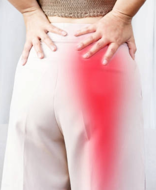
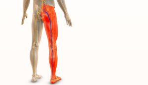
Radicular pain is caused by stimulation of the sensory root or dorsal root ganglion (DRG) of a spinal nerve (Merskey et al., 2007). Radicular pain differs from nociceptive pain in that it is caused by neural activity in the dorsal root rather than stimulation of peripheral nerve ends. As a result, it is distinct from both somatic pain and somatic-referred pain. Furthermore, radicular discomfort is not synonymous with radiculopathy. While radicular discomfort results from the formation of ectopic impulses, radiculopathy is caused by a blockage in the conduction of sensory and motor axons, resulting in nerve function loss (Merskey et al., 2007).
Mechanistically, radicular pain involves the spreading of afferent nociceptive input alongside intricate cellular and molecular processes that trigger and sustain heightened nociceptive signals. Anatomical (Aoki et al., 1999; Chen et al., 2007) and electrophysiological (Gosselin et al., 2010) studies indicate that radicular pain spreads to adjacent spinal segments. This explains the clinical observation of multiple nerve roots being affected despite a single pathology level. Although the propagation of radicular afferent signals is complex, a clear sequence in the inflammatory cascade is evident (Bennett et al., 2014). The process starts with the degeneration of the disc nerve, releasing proinflammatory cytokines both at the lesion site and distally through Wallerian degeneration (WD). During the distal axon degeneration via WD, TNF-α is released by Schwann cells, endothelial cells, mast cells, and resident macrophages at the injury site. This leads to ectopic firing at the dorsal root ganglion (DRG), which then increases the release of neurotrophins and ectopic firing at the dorsal horn, ultimately resulting in central sensitization (Cunha et al., 2003; Nakamura et al., 2004; Sabin et al., 1990; Olmarker et al., 1999). The cellular-molecular cascade can also originate from herniated disc material. When material from the nucleus pulposus extrudes onto the spinal nerve, it leads to edema and ischemia. Experimental studies have shown that exposure of the nerve root near the DRG to nucleus pulposus material increases endoneurial fluid pressure and reduces blood flow to the DRG, causing secondary edema (Olmarker et al., 1998). A crucial mediator released following disc herniation is the proinflammatory cytokine TNF-α. When TNF-α reaches the nerve root, it triggers the production of the neurotrophic factor (NGF) in the surrounding inflamed tissue (Olmarker et al., 1997; Ozaktay et al., 2003; Takebayashi et al., 2003). This process then induces the production of another neurotrophin, brain-derived neurotrophic factor (BDNF), in the DRG (Takebayashi et al., 2003; Zhang et al., 2012). Beyond their neurotrophic properties, such as promoting nerve sprouting, both NGF and BDNF are crucial in the development of central sensitization (Fukuoka et al., 2006) and play significant roles in the pathophysiology of radicular pain. Additionally, the release of cytokines can directly affect the activity and expression of various ion channels in the DRG (Vollmer et al., 2009). In radicular pain, ectopic discharges can occur at various locations within the nervous system. Electrophysiological studies estimate that about 75% of these discharges originate in the DRG, with only 25% occurring at the lesion or neuroma (Devor et al., 2000). Cytokines and neurotrophins can trigger the modulation and phosphorylation of ion channels, leading to the generation of ectopic action potentials or ectopic firing (Burchiel et al., 1994; Kovacs et al., 2011). The repetitive firing of pain afferents is a key physiological factor in central sensitization (Nakamura et al., 2004), significantly contributing to the clinical manifestation of radicular pain (Bennett et al., 2014).
Conservative and Surgical Approaches to Treating Radicular Pain
For managing radicular pain, healthcare providers often recommend “conservative therapy,” which typically includes exercises, traction, analgesics, and other measures like cervical collars for neck-related radicular pain. However, multiple studies have shown that these interventions do not offer benefits beyond the natural progression of the condition (Bogduk and Govind, 1999; Bogduk, 1999).
Research on physiotherapeutic treatments for radicular pain predominantly focuses on stabilization and motor control exercises or neural tissue management, with fewer studies exploring other techniques. Four trials with a low risk of bias (Ferreira et al., 2016; Hahne et al., 2017; Luijsterburg et al., 2008; Nee et al., 2012) were reviewed. Two studies (Nee et al., 2012; Ferreira et al., 2016) indicated that neural mobilizations might improve pain more than minimal care, though the confidence intervals reported included a wide range of possible effects. Conversely, Hahne et al. (2017) found no clinically meaningful effect when comparing individualized functional restoration programs, including stabilization exercises, to minimal care. Similarly, Luijsterburg et al. (2008) found no significant benefit when comparing general functional exercises to standard general practitioner care. Overall, it appears that interventions aimed at reducing neural mechanosensitivity might be more effective in alleviating radicular pain than those focused on spinal control and stability. However, this conclusion is based on indirect comparisons, and the clinical significance of the potential benefits remains uncertain. The mixed quality of trials and their conflicting results prevent drawing strong conclusions. While there is evidence that physiotherapy treatments can provide meaningful improvements in pain, quality of life, and disability for people with peripheral neuropathies and neuropathic pain, some high-quality studies do not support this. On a positive note, physiotherapeutic treatments are generally safe for people with radicular pain, allowing them to engage in exercise and enjoy its social, recreational, and health benefits. This is especially important for cancer patients, for whom exercise is strongly recommended due to its well-proven benefits beyond pain reduction (Mina et al., 2018). Nevertheless, regarding the benefits of exercise for peripheral neuropathic pain, physiotherapists should manage expectations realistically. The field may also benefit from promising results in preclinical sciences. A substantial body of preclinical literature suggests that exercise is not only hypoalgesic but also neuroprotective and neuroregenerative following focal and systemic nerve injury. The two main types of exercises examined are aerobic (e.g., running and swimming) and passive neurodynamic treatments. Aerobic exercise has been shown to reverse established neuropathic pain (Guo et al., 2019) and prevent its development (Grace et al., 2016). The neuroprotective benefits of aerobic exercise seem to be influenced more by variables such as exercise dosage than by the type of exercise (e.g., swimming vs. running). Low-to-moderate intensity exercise is neuroprotective, while moderate-to-high intensity exercise can be neurotoxic (Cobianchi et al., 2017; Cooper et al., 2016). Timing is also crucial; in rodent studies, exercising as soon as a week after nerve injury is more beneficial than delayed onset (Lopez-Alvarez et al., 2018). Similar to aerobic exercise, neurodynamic exercises and joint mobilizations appear to induce hypoalgesia and improve nerve regeneration in preclinical models of neuropathic pain, although most studies have focused on focal nerve injury (e.g., sciatic nerve injury) (Giardini et al., 2017; Martins et al., 2011; Santos et al., 2014; Santos et al., 2012). Only two trials provided detailed descriptions of adverse event definitions and recordings. Both indicated that mild adverse events were common. Nee et al. (2012) reported that 42% of patients experienced an adverse event, mainly neck or arm pain, which subsided within 24 hours in 95% of cases. Fritz et al. (2014) found that 56% of participants experienced adverse events, mostly worsening neck pain, with no difference in adverse event rates among the three trial arms. No serious or lasting adverse events were recorded.
Traditionally, surgery has been the primary treatment for radicular pain. Procedures such as laminectomy and microdiscectomy are commonly used for lumbar radicular pain, while foraminotomy is used for cervical radicular pain. Although studies indicate that surgery does not yield better long-term outcomes than conservative therapy, it has the notable advantage of providing immediate relief for patients with severe, unresponsive pain (Bogduk and Govind, 1999; Bogduk, 1999). Other interventions explored and recommended for radicular pain include epidural steroid injections and transforaminal steroid injections.
Conclusion
Radicular pain arises from sensory root or dorsal root ganglion (DRG) stimulation, distinct from nociceptive pain. It involves intricate cellular and molecular processes, leading to heightened nociceptive signaling and central sensitization. Conservative therapies, including exercises and analgesics, offer limited benefits beyond the condition’s natural progression. Physiotherapeutic interventions focusing on reducing neural mechanosensitivity may be more promising but lack direct comparison and clear clinical significance. Preclinical studies suggest exercise’s hypoalgesic, neuroprotective, and neuroregenerative effects, providing potential avenues for future research.
Learning Points
- Radicular pain results from sensory root or DRG stimulation, distinct from nociceptive pain.
- Conservative therapies offer limited benefits, with physiotherapy interventions targeting neural mechanosensitivity showing potential.
- Preclinical studies suggest exercise’s hypoalgesic, neuroprotective, and neuroregenerative effects, indicating promising avenues for future research.
- Adverse events from treatments are common but mostly mild and transient.
- Surgery provides immediate relief for severe, unresponsive pain but does not yield better long-term outcomes than conservative therapy.
References
- Aoki, Y., Rydevik, B. L., Kikuchi, S., & Olmarker, K. (1999). Local application of disc-related cytokines on spinal nerve roots. Spine, 24(9), 915-920.
- Bennett, G. J., Xie, Y. K., & Wang, X. M. (2014). Mechanisms of neuropathic pain. Molecular Pain, 10, 23.
- Bogduk, N. (1999). Medical Management of Acute Cervical Radicular Pain: An Evidence-Based Approach. Newcastle Bone and Joint Institute, Newcastle.
- Bogduk, N., & Govind, J. (1999). Medical Management of Acute Lumbar Radicular Pain: An Evidence-Based Approach. Newcastle Bone and Joint Institute, Newcastle.
- Burchiel, K. J. (1994). Effects of electrical and chemical stimulation on excitability of isolated human nerve. Pain, 58(3), 369-375.
- Chen, Y., Wu, W., Xu, X., Zhu, S., Yang, J., & Zhang, X. (2007). Pathogenesis of radicular pain in degenerative lumbar spinal stenosis. European Spine Journal, 16(9), 1373-1380.
- Cobianchi, S., Arbat-Plana, A., Lopez-Alvarez, V. M., & Navarro, X. (2017). Neuroprotective effects of exercise treatments after injury: the dual role of neurotrophic factors. Current Neuropharmacology, 15, 495–518.
- Cooper, M. A., Kluding, P. M., & Wright, D. E. (2016). Emerging relationships between exercise, sensory nerves, and neuropathic pain. Frontiers in Neuroscience, 10, 372.
- Cunha, F. Q., Poole, S., Lorenzetti, B. B., & Ferreira, S. H. (2003). The pivotal role of tumour necrosis factor alpha in the development of inflammatory hyperalgesia. British Journal of Pharmacology, 104(3), 669-674.
- Devor, M., Govrin-Lippmann, R., & Angelides, K. (2000). Na+ channel immunolocalization in peripheral mammalian axons and changes following nerve injury and neuroma formation. Journal of Neuroscience, 13(5), 1976-1992.
- Ferreira, G., Stieven, F., Araujo, F., Wiebusch, M., Rosa, C., Plentz, R., & Silva, M. (2016). Neurodynamic treatment did not improve pain and disability at two weeks in patients with chronic nerve-related leg pain: a randomised trial. Journal of Physiotherapy, 62, 197–202.
- Fritz, J. M., Thackeray, A., Brennan, G. P., & Childs, J. D. (2014). Exercise only, exercise with mechanical traction, or exercise with over-door traction for patients with cervical radiculopathy, with or without consideration of status on a previously described subgrouping rule: a randomized clinical trial. Journal of Orthopaedic & Sports Physical Therapy, 44, 45–57.
- Giardini, A. C., dos Santos, F. M., da Silva, J. T., de Oliveira, M. E., Martins, D. O., & Chacur, M. (2017). Neural mobilization treatment decreases glial cells and brain-derived neurotrophic factor expression in the central nervous system in rats with neuropathic pain induced by CCI. Pain Research & Management, 2017, 1–9.
- Grace, P. M., Fabisiak, T. J., Green-Fulgham, S. M., Anderson, N. D., Strand, K. A., Kwilasz, A. J., … & Watkins, L. R. (2016). Prior voluntary wheel running attenuates neuropathic pain. Pain, 157, 2012–23.
- Guo, J., Chen, B., Wang, Y., Zhu, Y., Song, G., Yang, Z., … & Chen, P. (2019). Meta-analysis of the effect of exercise on neuropathic pain induced by peripheral nerve injury in rat models. Frontiers in Neurology, 10, 636.
- Hahne, A. J., Ford, J. J., Hinman, R. S., Richards, M. C., Surkitt, L. D., Chan, A. Y. P., … & Taylor, N. F. (2017). Individualized functional restoration as an adjunct to advice for lumbar disc herniation with associated radiculopathy: A preplanned subgroup analysis of a randomized controlled trial. The Spine Journal, 17, 346–59.
- Kovacs, G. G., Laszlo, L., Kovacs, J., Trembeczki, K., Bogner, P., & Olah, I. (2011). Proteomics of cerebrospinal fluid in Alzheimer’s disease: Identification of biomarkers. Journal of Alzheimer’s Disease, 26(1), 269-278.
- Lopez-Alvarez, V. M., Puigdomenech, M., Navarro, X., & Cobianchi, S. (2018). Monoaminergic descending pathways contribute to modulation of neuropathic pain by increasing-intensity treadmill exercise after peripheral nerve injury. Experimental Neurology, 299, 42–55.
- Luijsterburg, P. A. J., Verhagen, A. P., Ostelo, R. W. J. G., van den Hoogen, H. J. M. M., Peul, W. C., Avezaat, C. J. J., & Koes, B. W. (2008). Physical therapy plus general practitioners’ care versus general practitioners’ care alone for sciatica: a randomised clinical trial with a 12-month follow-up. European Spine Journal, 17, 509–17.
- Martins, D. F., Mazzardo-Martins, L., Gadotti, V. M., Nascimento, F. P., Lima, D. A. N., Speckhann, B., … & Santos, A. R. S. (2011). Ankle joint mobilization reduces axonotmesis-induced neuropathic pain and glial activation in the spinal cord and enhances nerve regeneration in rats. Pain, 152, 2653–61.
- Mina, D. S., Langelier, D., Adams, S. C., Alibhai, S. M. H., Chasen, M., Campbell, K. L., … & Chang, E. (2018). Exercise as part of routine cancer care. The Lancet Oncology, 19, e433–6.
- Nee, R. J., Vicenzino, B., Jull, G. A., Cleland, J. A., & Coppieters, M. W. (2012). Neural tissue management provides immediate clinically relevant benefits without harmful effects for patients with nerve-related neck and arm pain: a randomised trial. Journal of Physiotherapy, 58, 23–31.
- Santos, F., Grecco, L., Pereira, M., Oliveira, M., Rocha, P., Silva, J., … & Chacur, M. (2014). The neural mobilization technique modulates the expression of endogenous opioids in the periaqueductal gray and improves muscle strength and mobility in rats with neuropathic pain. Behavioural Brain Function, 10, 19.
- Santos, F., Silva, J. T., Giardini, A. C., Rocha, P. A., Achermann, A. P., Alves, A. S., Britto, L. R., & Chacur, M. (2012). Neural mobilization reverses behavioral and cellular changes that characterize neuropathic pain in rats. Molecular Pain, 8, 57.
- Sabin, T. D., Mussell, E. R., Graham, M. K., & Burchiel, K. J. (1990). Effects of nucleus pulposus on peripheral nerve: a model of chemical radiculitis. Journal of Orthopaedic Research, 8(6), 843-853.
- Takebayashi, T., Cavanaugh, J. M., Kallakuri, S., Ozaktay, A. C., & Chen, C. (2003). Effect of nucleus pulposus on nerve root neural activity, mechanosensitivity, axonal morphology, and sodium channel expression. Spine, 28(1), 25-35.
- Vollmer, C., Tanelian, D. L., & Shelton, D. L. (2009). The role of cytokines in the modulation of nociceptive neuron excitability. Neuroscience, 58(2), 399-409.
- Zhang, J., Shi, X. Q., Echeverry, S., Mogil, J. S., De Koninck, Y., & Rivest, S. (2012). Expression of brain-derived neurotrophic factor in primary sensory neurons is stimulated by fractalkine and contributes to inflammatory pain. Journal of Pain, 13(4), 375-383.