A classification method based on symptom behaviour sub-grouping improves treatment outcomes, according to recent research (Fritz and George, 2000; Long et al., 2004). The association between the sacroiliac joint (SIJ) and low back pain has been a source of contention, with some researchers seeing SIJ pain as a key contribution to the problem and others dismissing it as inconsequential or irrelevant. The clinical reasoning technique utilised by physicians is characterised by the formation of a diagnosis based on a combination of findings.
SIJ pain prevalence:
The prevalence of SIJ diseases ranges from 13% to 53% (Dreyfuss et al., 1996; Maigne et al., 1996). It is widely acknowledged that approximately 13% of people with chronic low back pain have the SIJ as the source of their discomfort (Maigne et al., 1996).
SIJ biomechanical and anatomical considerations:
The SIJ’s bone anatomy, such as size and morphologic properties, varies between individuals, and its shape evolves from childhood to adulthood (Slipman CW, et al., 2001, Vleeming A, et Al., 2014). Only the anterior third of the SIJ’s articular surface is a synovial joint, with the rest of the articular surface made up of ligamentous connections (Harrison DE, et Al., 1997, Cohen SP, 2005). The SIJ’s movement was restricted by its anatomical nature ( Cho HJ, and Kwak DS., 2021). The SIJ has a limited range of motion, with less than 4° of rotation and up to 1.6 mm of translation (Sturesson B, et al., 1989, Sturesson B, et al., 2000). Sturesson et al. 1999 discovered no difference in range of motion between the symptomatic and asymptomatic sides in patients assumed to have a SIJ source of pain, which was also supported by a study conducted by Kibsgård TJ et al., who concluded that in patients with PGP, movements in the SIJs during the single-leg stance are small and almost undetectable, and they are also highlighted. The sacroiliac joint movement during single-leg stance is modest and nearly undetectable by precision radiostereometric measurement (Kibsgård TJ et al., 2014). There are also differences in pelvic morphology, which may have a considerable impact on measures of pelvic tilt and innominate rotational asymmetry (Preece SJ, et al., 2008).
Is it SIJ pain or SIJ dysfunction?
There are two clinical views to consider: the SIJ as a load-transferring mechanical connection between the pelvis and the spine that may create painful stimuli from the SIJ or other tissues, and the SIJ as a source of pain. The issue is that no widely accepted reference standard exists for SIJ deficiency (laslett M., 2008). As a result, tests for SIJ dysfunction have low inter-examiner reliability. Because there is no reference standard for SIJ dysfunction, the validity of the tests for this illness is uncertain. Inter-examiner reliability and therapeutically relevant validity against an appropriate reference standard are acceptable for tests that stress the SIJ in order to elicit familiar pain. It is unclear whether provocation testing can accurately identify extra-articular SIJ pain origins (Laslett M., 2008).
SIJ pain diagnosis:
SIJ pain and discogenic pain rarely coexist. provocation SIJ tests were frequently positive in patients with nerve root pain caused by a herniated lumbar disc, as well as in patients whose symptoms could be forced to centralise during a McKenzie-style physical examination. The phenomenon of centralization has been studied and tested for reliability and validity numerous times (Razmjou H, et al., 2000; Kilpikoski S, et al., 2002; Aina A, et al., 2004; Clare HA, et al., 2005; Donelson R, et al., 1990, 1997, 1991; Wetzelf T, Donelson R, 2003; Long A, et al., 2004; Donelson R, 2007; Werneke MW, et al., 1999, 2001, 2003, 2005). It has since been discovered to be extremely specific to discogenic pain and is not seen in individuals with confirmed SIJ or facet joint discomfort (Laslett M. et al., 2003; 2005, 2006). The absence of centralisation/peripheralisation renders the pathology of an intervertebral disc derangement implausible and increases the validity of the SIJ tests (Donelson et al., 1997; Laslett et al., 2003; Young et al., 2003).
On this premise, it appears plausible to conclude that SIJ tests that are positive in the presence of the centralization phenomenon are mistakenly positive. Restricting SIJ test interpretation to non-centralization cases raises the specificity of three or more positive pain provocation SIJ tests from 78% to 87% while maintaining sensitivity at 91% (Laslett M. et al., 2003). Other clinical connections studied include the fact that patients with SIJ pain at or above the level of L5 are uncommon, and the presence of midline pain tends to rule out the SIJ as a pain generator (Young et al., 2003). This clinical reasoning process can be thought of as a clinical prediction rule for identifying a subset of individuals who are most likely to experience SIJ pain.
Werneke and Hart (2003) found that a percentage of patients who do not exhibit centralisation at their initial appointment may do so at later visits. This is another major reason to evaluate only the lumbar spine to avoid missing this important prognostic finding. As a result, McKenzie and May (2003) advocate excluding the lumbar spine with a 24-48-hour activity trial before performing additional joint examination.
The following clinical prediction rule for sacroiliac joint pain can be simply used to the vast majority of back pain patients:
- A McKenzie examination of repeated movements or sustained positions does not result in pain centralization.
- The normal discomfort is elicited by three or more provocation tests.
In summary, Only after excluding lumbar and hip sources of pain should a determination be made that a painful sacroiliac joint lesion occurs. three or more positive pain provocation SIJ tests have 91% sensitivity and 78% specificity. The prevalence of three or more positive tests rises to 87% in patients whose symptoms do not migrate towards the spinal midline, i.e., centralise. Patients with three or more positive provocation SIJ tests and whose symptoms cannot be induced to centralise have a 77% chance of having SIJ pain in chronic back pain populations and an 89% chance in pregnant back pain populations. There were significant connections between SI joint pain provocation tests, pain while rising from a sitting position, unilateral discomfort, and a lack of midline lumbar pain and SI joint pain (Young S. et al., 2003).
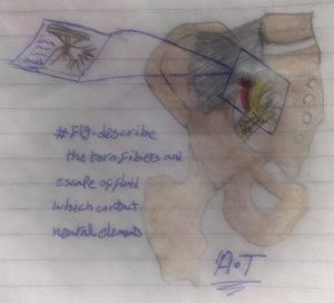
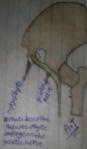
Communication channels between the SIJ and adjacent neural structures (Fortin JD et al., 1999):
- Synovial fluid leakage from a neighbouring joint may lead to radicular symptoms.
- Osteophytes in the sacroiliac joints are most commonly found in the anteroinferior area of the joint [in females] and, if large enough, can cause sciatic nerve root compression.
Extra-spinal diseases that cause sciatica are “rare.” However, failure to identify these leads to incorrect diagnosis and management, resulting in poor outcomes.
SIJ pain relief:
SIJ causes discomfort for one of two reasons:
- There is some evidence that an inflammatory condition within the joint is causing or contributing to the pain (Heuft-Dorenbosch L, et al., 2006; Slipman CW, et al., 1996).
- The joint is unstable due to ligamentous laxity or joint capsule tears (Fortin JD, et al., 1994; Fortin JD, et al., 1996; Berthelot JM, et al., 2006; van Wingerden JP, et al., 2000).
So, how do we treat people who are at high risk of SIJ pain?
Exercises targeted at improving lumbopelvic stability and intra-articular steroid injections are the treatments with the greatest promise for pain and disability reduction.
- The manipulation technique affects the lumbar spine as well as the SI area (Flynn T. et al., 2003).
- Injections of corticosteroids (Pereira PL et al., 2000; Maugars Y et al., 1996; Slipman CW et al., 2001) and phenol injections (Ward S. et al., 2002) are other procedures not available to physical therapists that may be effective in the treatment of persistent SIJ discomfort. They are less invasive and appear to be effective in a subset of SIJ pain patients, particularly when imaging evidence of sacroiliitis is present, according to Burnham RS, Yasui Y, 2007; Vallejo R, et al., 2006; Ferrante FM, et al., 2001; Yin W, et al., 2003; Cohen SP, Abdi S, 2003.
- Surgical debridement (Haufe SM, Mork A., 2005) and fusion (Slipman CW, et al., 1998) are more invasive, but they appear to offer a moderate probability of pain reduction and functional improvement in patients with proven SIJ pain who have failed to respond to more conservative therapies.
References:
- Berthelot JM, Labat JJ, Le Goff B, Gouin F, Maugers Y. Provocative sacroiliac joint maneuvers and sacroiliac joint block are unreliable for diagnosing sacroiliac joint pain. Joint Bone Spine 2006;73:17–23.
- Burnham RS, Yasui Y. An alternate method of radiofrequency neurotomy of the sacroiliac joint: A pilot study of the eff ect on pain, function, and satisfaction. Reg Anesth Pain Med 2007;32:12–19.
- Cho HJ, Kwak DS. Movement of the sacroiliac joint: Anatomy, systematic review, and biomechanical considerations. Proc Inst Mech Eng H. 2021 Mar;235(3):357-364.
- Cohen SP, Abdi S. Lateral branch blocks as a treatment for sacroiliac joint pain: A pilot study. Reg Anesth Pain Med 2003;28:113– 119.
- Cohen SP. Sacroiliac joint pain: a comprehensive review of anatomy, diagnosis, and treatment. Anesth Analg 2005; 101: 1440–1453.
- Donelson R, Aprill C, Medcalf R, Grant W. A prospective study of centralisation of lumbar and referred pain. A predictor of symptomatic discs and annular competence. Spine 1997; 22:1115–22.
- Ferrante FM, King LF, Roche EA, et al. Radiofrequency sacroiliac joint denervation for sacroiliac syndrome. Regl Anesth Pain Med 2001;26:137–142.
- Flynn T, Fritz JM, Whitman J, et al. A clinical prediction rule for classifying patients with low back pain who demonstrate short-term improvement with spinal manipulation. Spine 2003;27:2835–2843.
- Fortin JD, Aprill C, Pontieux RT, Pier J. Sacroiliac joint: Pain referral maps upon applying a new injection/arthrography technique. Part II: Clinical evaluation. Spine 1994;19:1483–1489.
- Fortin JD, Washington WJ, Falco FJE. Three pathways between the sacro-iliac joint and neural structures. AJNR 1999;20:1429– 1434.
- Fritz JM, George S. The use of a classification approach to identify subgroups of patients with acute low back pain. Spine 2000;25:106–14.
- Haufe SM, Mork AR. Sacroiliac joint debridement: A novel technique for the treatment of sacroiliac joint pain. Photomed Laser Surg 2005;23:596–598.
- Harrison DE, Harrison DD and Troyanovich SJ. The sacroiliac joint: a review of anatomy and biomechanics with clinical implications. J Manipulative Physiol Ther 1997; 20: 607–617.
- Heuft -Dorenbosch L, Weijers R, Landewe R, van der Linden S, van der Heijde D. Magnetic resonance imaging changes of sacroiliac joints in patients with recent-onset infl ammatory back pain: Inter-reader reliability and prevalence of abnormalities. Arthritis Res Ther 2006;8:R11.
- Kibsgård TJ, Røise O, Sturesson B, Röhrl SM, Stuge B. Radiosteriometric analysis of movement in the sacroiliac joint during a single-leg stance in patients with long-lasting pelvic girdle pain. Clin Biomech. 2014Apr;29(4):406-11.
- Laslett M. Evidence-based diagnosis and treatment of the painful sacroiliac joint. J Man Manip Ther. 2008;16(3):142-52.
- Laslett M, Young S, Aprill CN, McDonald B. Diagnosing painful sacroiliac joints: a validity study of a McKenzie evaluation and sacroiliac provocation tests. Australian Journal of Physiotherapy 2003;46:89–97.
- Long A, Donelson R, Fung T. Does it matter which exercise? Spine 2004;29(23):2593–602.
- Maugars Y, Mathis C, Berthelot JM, Charlier C, Prost A. Assessment of the effi cacy of sacroiliac corticosteroid injections in spondylarthropathies: A double-blind study. Br J Rheumatol 1996;35:767–770.
- McKenzie RA, May S. Mechanical diagnosis and therapy: the lumbar spine. 2nd ed. Waikanae: Spinal Publications; 2003.
- Pereira PL, Gunaydin I, Trubenbach J, et al. Interventional MR imaging for injection of sacroiliac joints in patients with sacroiliitis. AJR Am J Roentgenol 2000;175:265– 266.
- Preece SJ, Willan P, Nester CJ, Graham-Smith P, Herrington L, Bowker P. Variation in pelvic morphology may prevent the identification of anterior pelvic tilt. J Man Manip Ther. 2008;16(2):113-7.
- Slipman CW, Sterenfeld EB, Chou LH, Herzog R, Vresilovic E. Th e value of radionuclide imaging in the diagnosis of sacroiliac joint syndrome. Spine 1996;21:2251–2254.
- Slipman CW, Sterenfeld EB, Chou LH, Herzog R, Vresilovic E. Th e predictive value of provocative sacroiliac joint stress maneuvers in the diagnosis of sacroiliac joint syndrome. Arch Phys Med Rehabil 1998;79:288.
- Slipman CW, Lipetz JS, Plastaras CT, et al. Fluoroscopically guided therapeutic sacroiliac joint injections for sacroiliac joint syndrome. Am J Phys Med Rehabil 2001;80:425– 432.
- Slipman CW, Whyte WS 2nd, Chow DW, et al. Sacroiliac joint syndrome. Pain Physician 2001; 4: 143–152.
- Sturesson B, Selvik G, Uden A. Movements of the sacroiliac joints: A roentgnen stereophotogrammetric analysis. Spine 1989;14:162–165.
- Sturesson B, Uden A, Vleeming A. A radiostereometric analysis of the movements of the sacroiliac joints in the reciprocal straddle position. Spine 2000;25:214–217.
- Sturesson B. Load and movement of the sacroiliac joint. PhD thesis, Lund University, Malmo, Sweden,1999;29–35.
- Vallejo R, Benyamin RM, Kramer J, Stanton G, Joseph NJ. Pulsed radiofrequency denervation for the treatment of sacroiliac joint syndrome. Pain Med 2006;7:429–434.
- van Wingerden JP, Vleeming A, Buyruk HM, Raissadat K. Stabilization of the sacroiliac joint in vivo: Verification of muscular contribution to force closure of the pelvis. Eur Spine J 2004;13:199–205.
- Vleeming A, Schuenke MD, Masi AT, et al. The sacroiliac joint: an overview of its anatomy, function and potential clinical implications. J Anat 2012; 221: 537– 567.
- Ward S, Jenson M, Royal MA, Movva V, Bhakta B, Gunyea I. Fluoroscopy-guideds acroiliac joint injections with phenol ablation for persistent sacroiliitis: A case series.Pain Pract 2002;2:332–335.
- Werneke M, Hart DL. Discriminant validity and relative precision for classifying patients with non-specific neck and back pain by anatomic pain patterns. Spine 2003;28:161–6.
- Yin W, Willard F, Carreiro J, Dreyfuss P. Sensory stimulation-guided sacroiliac joint radiofrequency neurotomy: Technique based on neuroanatomy of the dorsal sacral plexus.Spine 2003;28:2419–2425.
- Young S, Aprill CN, Laslett M. Correlation of clinical examination characteristics with three sources of chronic low back pain. The Spine Journal 2003;3:460–5.