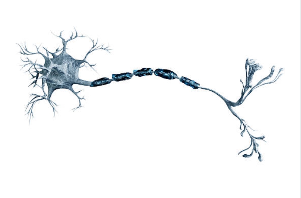
This blog summarises the available evidence on the influence of entrapment neuropathies on the anatomical and physiological features of the peripheral nervous system that have previously been discussed. Let’s get started!
Entrapment Neuropathies and Ischaemia
Entrapment neuropathies are hypothesised to disrupt intraneural blood flow by reversing the pressure gradient required for optimal blood supply. Extraneural pressures as low as 20–30 mmHg interrupt intraneural venous circulation, while pressures as high as 40–50 mmHg decrease arteriolar and capillary blood flow (Rydevik B. et al. 1981).
Extraneural pressure is higher in patients with entrapment neuropathies. Pressures above 50 mmHg were measured around the afflicted nerve roots in patients with lumbar disc herniations, with some patients exhibiting pressures as high as 250 mmHg (Takahashi K. et al. 1999). Such increased pressures, especially if present for an extended period of time, will be sufficient to reverse the usual pressure gradient, creating venous return obstruction with subsequent intraneural circulation slowing and oedema formation (Sunderland S. 1976).
Patients with entrapment neuropathies frequently have transient ischaemia. It not only explains the traditional position-dependent paraesthesia, but it can also help reproduce symptoms during provocative exercises. Whereas real neurological abnormalities are usually persistent, fluctuations with positional changes, for example, have been observed (Hough AD et al., 2007; Sabbahi MA; Ovak-Bittar F., 2018). Such fluctuations may be due to ischemic conduction block rather than demyelinating conduction block.
Ischemia can also cause the normal nighttime aggravation of symptoms that subsides with modest movement. The physiological nightly decline in blood pressure and associated dip in intraneural blood flow (Low PA, Tuck RR. 1984) can reverse the pressure gradient, resulting in ischaemia, alterations in metabolic activity, and ectopic firing. Venous distension can potentially contribute to pain by stimulating venous afferents (Sumikawa K. et al. 2001). Ischemia may be seen in mild entrapment neuropathies in the absence of structural abnormalities. This is demonstrated by the quick alleviation of symptoms in certain patients following surgery (Werner RA; Andary M. 2002). Enlargement of the compressed nerves (Nakamichi KI, Tachibana S. 2000; Yoon JS, et al. 2010) and increases in signal intensity on specialised magnetic resonance sequences (Cudlip SA, et al. 2002; Sirvanci M, et al. 2009; Lewis AM, et al. 2010) are clinical signs of oedema in entrapment neuropathies.
Oedema can eventually lead to intraneural and extraneural fibrotic alterations affecting connective and adipose tissues if left untreated (Mackinnon SE, 2002). Neural fibrosis has been reported in both radiculopathies (Ido K., Urushidani H. 2001) and peripheral nerve trunk entrapments (e.g., cubital and carpal tunnel syndrome) (Abzug JM et al. 2012).
Extraneural fibrotic alterations may be responsible for compressed nerves’ reduced gliding ability in relation to their surrounding tissues. However, such biomechanical changes are unlikely to be the only mechanism underlying signs of increased nerve sensitivity during neurodynamic testing (for example, provocation of symptoms, change in symptoms by moving joints away from the symptomatic area [structural differentiation], and potentially reduced range of motion). Positive neurodynamic tests in individuals with entrapment neuropathies may instead be explained by a variety of neurophysiological alterations leading to greater neural mechanosensitivity.
Entrapment Neuropathies Cause Demyelination
Focal demyelination is thought to be a defining feature of nerve entrapments (Mackinnon SE, 2002). Demyelination is a common side effect of prolonged ischaemia, which causes Schwann cell failure (Nukada H, et al. 1993), but it can also be linked to mechanical deformation (Lin MY, et al. 2012) or a cytotoxic environment caused by processes such as inflammation.
Histological data from animal models of mild nerve compression (O’Brien JP, et al. 1987; Schmid AB, et al. 2012; Gupta R, et al. 2004) and from patients with entrapment neuropathies (Foix C, Marie P. 1913; Neary D. 1975; Mackinnon SE, et al. 1986) confirm focal demyelination and remyelination with intra-fascicular fibrosis and connective tissue thickening. Similar histological results have been found in asymptomatic people at common entrapment locations (Jefferson D., Eames RA. 1979; Neary D., et al. 1975). This shows that such focal histological changes may not always result in symptoms. Myelin alterations spread beyond the lesion site in addition to focal demyelination. Demyelination of the tibial nerve following focal mild nerve compression of the sciatic nerve in rats (Schmid AB, et al. 2013) and the presence of elongated nodes of Ranvier in skin biopsies taken more than 9 cm beyond the compression site in patients with carpal tunnel syndrome (Schmid AB, et al. 2012) support this.
Because ion channels can be more easily inserted into the membrane at demyelinated regions and the neuronal cell body, changes in ion channel configuration are more common at these sites (Liu X, et al., 2001; Jiang YQ, et al., 2008; Drummond PD, 2014; England JD, et al., 1994).
These ion channel alterations have been linked to spontaneous ectopic action potential production (Amir R. et al., 1999; Chen Y., Devor M., 1998). Because action potentials are normally relayed but do not originate along the axon or cell body, these sites are referred to as aberrant impulse-producing sites.
Changes in the configuration of ion channels in entrapment neuropathies may not only underpin ectopic activity (e.g., paraesthesia, Tinel’s sign, nerve mechanosensitivity upon palpation), but may also impair normal saltatory impulse conduction, resulting in the characteristic slowing or block of nerve conduction during electrodiagnostic testing (Mallik A., Weir AI., 2005; Kiernan MC, et al., 1999).
Entrapment Neuropathies Affect Both Large- and Small-Diameter Nerve Fibres
In an animal model of mild nerve compression (Schmid AB, et al. 2013), a predominant compromise of small axons with structural sparing of large axons (apart from demyelination) was observed, contrary to common beliefs that small fibres are relatively resistant to compression (Dahlin LB, et al. 1989).
Most studies using quantitative sensory testing suggest loss of function of small myelinated and unmyelinated fibres (deficit in cold and warm detection) in both lumbar and cervical radiculopathy as well as CTS (Schmid AB, et al. 2014; Witt JC, et al. 2004; Tamburin S, et al. 2010; Chien A, et al. 2008; Tampin B, et al. 2012; Samuelsson L, Lundin A. 2002).
Several studies find significant alterations of sympathetic axon function in patients with CTS and radiculopathy (Wilder-Smith EP, et al. 2003; Kiylioglu N., 2007; Kuwabara S, et al. 2008; Kiylioglu N, et al. 2005; Reddeppa S, et al. 2000; Erdem Tilki H, et al. 2014), and laser-evoked brain potentials (mediated by A and C fibres) are reduced in patients with CTS (Arendt-Nielsen L, et al. 1991).
In patients with entrapment neuropathies, tiny fibres are damaged not only in function but also in structural integrity (Schmid AB et al. 2014; Ramieri G et al. 1995). This is evidenced by a significant loss of epidermal nerve fibres in the skin of CTS sufferers.
The effect of the detected loss of tiny fibres on the diagnosis and management of entrapment neuropathies has to be investigated further. Interestingly, preliminary studies in patients with CTS symptoms but normal neurophysiology imply that alterations in electrodiagnostic testing may precede changes in small axon dysfunction or loss (Schmid AB et al., 2024; Tamburin S et al., 2010). These findings suggest that tests for small fibre function should be included in the early diagnosis of entrapment neuropathies. Simple bedside neurological tests (e.g., pin prick sensitivity, cold and warm detection), as well as more equipment-intensive examinations such as quantitative sensory testing, sympathetic reflex testing, laser or heat-evoked brain potentials, or skin biopsies, are examples of clinical small fibre tests.
References
- Abzug JM, Jacoby SM, Osterman AL. Surgical options for recalcitrant carpal tunnel syndrome with perineural fibrosis. Hand (NY) 2012;7(1):23–9.
- Amir R, Michaelis M, Devor M. Membrane potential oscillations in dorsal root ganglion neurons: role in normal electrogenesis and
- neuropathic pain. J Neurosci 1999;19(19):8589–96.
- Arendt-Nielsen L, Gregersen H, Toft E, et al. Involvement of thin afferents in carpal tunnel syndrome: evaluated quantitatively by argon laser stimulation. Muscle Nerve 1991;14(6):508–14.
- Chen Y, Devor M. Ectopic mechanosensitivity in injured sensory axons arises from the site of spontaneous electrogenesis. Eur J Pain 1998;2(2):165–78.
- Chien A, Eliav E, Sterling M. Whiplash (grade II) and cervical radiculopathy share a similar sensory presentation: an investigation using quantitative sensory testing. Clin J Pain 2008; 24(7):595–603.
- Cudlip SA, Howe FA, Clifton A, et al. Magnetic resonance neurography studies of the median nerve before and after carpal tunnel decompression. J Neurosurg 2002;96(6):1046–51.
- Dahlin LB, Shyu BC, Danielsen N, et al. Effects of nerve compression or ischaemia on conduction properties of myelinated and non-myelinated nerve fibres. An experimental study in the rabbit common peroneal nerve. Acta Physiol Scand 1989;136(1): 97–105.
- Drummond PD, Drummond ES, Dawson LF, et al. Upregulation of alpha1-adrenoceptors on cutaneous nerve fibres after partial sciatic nerve ligation and in complex regional pain syndrome type II. Pain 2014;155(3):606–16.
- England JD, Gamboni F, Ferguson MA, et al. Sodium channels accumulate at the tips of injured axons. Muscle Nerve 1994;17(6): 593–8.
- Erdem Tilki H, Coskun M, Unal Akdemir N, et al. Axon count and sympathetic skin responses in lumbosacral radiculopathy. J Clin Neurol 2014;10(1):10–16.
- Foix C, Marie P. Atrophie isolée de l’éminence thénar d’origin névritique. Rôle du ligament annulaire antérieur du carpe dans la pathogénie de la lésion. Rev Neurol 1913;26:647–8.
- Gupta R, Rowshan K, Chao T, et al. Chronic nerve compression induces local demyelination and remyelination in a rat model of carpal tunnel syndrome. Exp Neurol 2004;187(2):500–8.
- Hough AD, Moore AP, Jones MP. Reduced longitudinal excursion of the median nerve in carpal tunnel syndrome. Arch Phys Med Rehabil 2007;88(5):569–76.
- Ido K, Urushidani H. Fibrous adhesive entrapment of lumbosacral nerve roots as a cause of sciatica. Spinal Cord 2001;39(5):269–73.
- Jefferson D, Eames RA. Subclinical entrapment of the lateral femoral cutaneous nerve: an autopsy study. Muscle Nerve 1979;2(2):145–54.
- Jiang YQ, Xing GG, Wang SL, et al. Axonal accumulation of hyperpolarization-activated cyclic nucleotide-gated cation channels contributes to mechanical allodynia after peripheral nerve injury in rat. Pain 2008;137(3):495–506.
- Kiernan MC, Mogyoros I, Burke D. Conduction block in carpal tunnel syndrome. Brain 1999;122(5):933–41.
- Kiylioglu N, Akyol A, Guney E, et al. Sympathetic skin response in idiopathic and diabetic carpal tunnel syndrome. Clin Neurol Neurosurg 2005;108(1):1–7.
- Kiylioglu N. Sympathetic skin response and axon counting in carpal tunnel syndrome. J Clin Neurophysiol 2007;24(5):424, author reply.
- Kuwabara S, Tamura N, Yamanaka Y, et al. Sympathetic sweat responses and skin vasomotor reflexes in carpal tunnel syndrome. Clin Neurol Neurosurg 2008;110(7):691–5.
- Lewis AM, Layzer R, Engstrom JW, et al. Magnetic resonance neurography in extraspinal sciatica. Arch Neurol 2006;63(10): 1469–72.
- Lin MY, Frieboes LS, Forootan M, et al. Biophysical stimulation induces demyelination via an integrin-dependent mechanism. Ann Neurol 2012;72(1):112–23.
- Liu X, Zhou JL, Chung K, et al. Ion channels associated with the ectopic discharges generated after segmental spinal nerve injury in the rat. Brain Res 2001;900(1):119–27.
- Low PA, Tuck RR. Effects of changes of blood pressure, respiratory acidosis and hypoxia on blood flow in the sciatic nerve of the rat. J Physiol 1984;347:513–24.
- Mackinnon SE, Dellon AL, Hudson AR, et al. Chronic human nerve compression–a histological assessment. Neuropathol Appl Neurobiol 1986;12(6):547–65.
- Mackinnon SE. Pathophysiology of nerve compression. Hand Clin 2002;18(2):231–41.
- Mallik A, Weir AI. Nerve conduction studies: essentials and pitfalls in practice. J Neurol Neurosurg Psychiatry 2005;76(Suppl. 2):ii23–31.
- Nakamichi KI, Tachibana S. Enlarged median nerve in idiopathic carpal tunnel syndrome. Muscle Nerve 2000;23(11):1713–18.
- Neary D, Ochoa J, Gilliatt RW. Sub-clinical entrapment neuropathy in man. J Neurol Sci 1975;24(3):283–98.
- Neary D. The pathology of ulnar nerve compression in men. Neuropathol Appl Neurobiol 1975;1:69–88.
- Nukada H, Powell HC, Myers RR. Spatial distribution of nerve injury after occlusion of individual major vessels in rat sciatic nerves. J Neuropathol Exp Neurol 1993;52(5):452–9.
- O’Brien JP, Mackinnon SE, MacLean AR, et al. A model of chronic nerve compression in the rat. Ann Plast Surg 1987; 19(5):430–5.
- Papanicolaou GD, McCabe SJ, Firrell J. The prevalence and characteristics of nerve compression symptoms in the general population. J Hand Surg [Am] 2001;26(3):460–6
- Ramieri G, Stella M, Calcagni M, et al. An immunohistochemical study on cutaneous sensory receptors after chronic median nerve compression in man. Acta Anat 1995;152(3):224–9.
- Reddeppa S, Bulusu K, Chand PR, et al. The sympathetic skin response in carpal tunnel syndrome. Auton Neurosci 2000; 84(3):119–21.
- Rydevik B, Lundborg G, Bagge U. Effects of graded compression on intraneural blood blow. An in vivo study on rabbit tibial nerve. J Hand Surg 1981;6(1):3–12.
- Sabbahi MA, Ovak-Bittar F. Electrodiagnosis-based management of patients with radiculopathy: the concept and application involving a patient with a large lumbosacral disc herniation. Clin Neurophysiol Pract 2018;3:141–7.
- Samuelsson L, Lundin A. Thermal quantitative sensory testing in lumbar disc herniation. Eur Spine J 2002;11(1):71–5.
- Schmid AB, Coppieters MW, Ruitenberg MJ, et al. Local and remote immune-mediated inflammation after mild peripheral nerve compression in rats. J Neuropathol Exp Neurol 2013; 72(7):662–80.
- Schmid AB, Coppieters MW, Ruitenberg MJ, et al. Local and remote immune-mediated inflammation after mild peripheral nerve compression in rats. J Neuropathol Exp Neurol 2013; 72(7):662–80.
- Schmid AB, Bland JD, Bhat MA, et al. The relationship of nerve fibre pathology to sensory function in entrapment neuropathy. Brain 2014;137(Pt 12):3186–99.
- Schmid AB, Bland JD, Bhat MA, et al. The relationship of nerve fibre pathology to sensory function in entrapment neuropathy. Brain 2014;137(Pt 12):3186–99.
- Sunderland S. The nerve lesion in the carpal tunnel syndrome. J Neurol Neurosurg Psychiatry 1976;39(7):615–26.
- Sirvanci M, Kara B, Duran C, et al. Value of perineural edema/ inflammation detected by fat saturation sequences in lumbar magnetic resonance imaging of patients with unilateral sciatica. Acta Radiol 2009;50(2):205–11.
- Sumikawa K, Sakai T, Ono T. Peripheral vascular pain. Nippon Rinsho 2001;59(9):1733–7.
- Takahashi K, Shima I, Porter RW. Nerve root pressure in lumbar disc herniation. Spine 1999;24(19):2003–6.
- Tamburin S, Cacciatori C, Praitano ML, et al. Median nerve small- and large-fiber damage in carpal tunnel syndrome: a quantitative sensory testing study. J Pain 2010;12(2):205–12.
- Tampin B, Slater H, Hall T, et al. Quantitative sensory testing somatosensory profiles in patients with cervical radiculopathy are distinct from those in patients with nonspecific neck-arm pain. Pain 2012;153(12):2403–14.
- Werner RA, Andary M. Carpal tunnel syndrome: pathophysiology and clinical neurophysiology. Clin Neurophysiol 2002;113(9): 1373–81.
- Wilder-Smith EP, Fook-Chong S, Chew SE, et al. Vasomotor dysfunction in carpal tunnel syndrome. Muscle Nerve 2003; 28(5):582–6.
- Witt JC, Hentz JG, Stevens JC. Carpal tunnel syndrome with normal nerve conduction studies. Muscle Nerve 2004;29(4): 515–22.
- Yoon JS, Walker FO, Cartwright MS. Ulnar neuropathy with normal electrodiagnosis and abnormal nerve ultrasound. Arch Phys Med Rehabil 2010;91(2):318–20.
