In Western industrialised countries, low-back pain (LBP) is a common cause of disability. Although many people have at least one episode of low-back pain throughout their lives, no identifiable disease is found in up to 85% of patients (Deyo et al. 1992). Low back-related leg pain, which accounts for 23% to 57% of all LBP cases (Scharfer A, et al. 2007). In patients who report symptoms radiating into the leg (spine-related leg pain), clinicians evaluate the possible causes of radiculopathy (compression of the nerve root) through history and physical examination. Part of These process is clinical neurological tests include sensory, motor, reflex, neurodynamic and nerve trunk palpation procedures designed to assess the physiological and bio-mechanical status of specific lumbar nerve roots thought to be responsible for the patient’s signs and symptoms (Jensen MC, et al. 1994). Clinically, the definition of lumbo-sacral radiculopathy is “objective loss of sensory and motor function with or without accompanied spinal and/or referred leg pain following a mechanical or biochemical dysfunction of lumbar and sacral spinal nerve roots and their associated dorsal root ganglions (DRGs)”. As a result, the purpose of this blog was to determine the accuracy of clinical neurological testing in identifying lumbo-sacral radiculopathy.
Neurological Integrity Tests
As previously stated, practitioners have used neurological integrity tests to determine lumbosacral radiculopathy for many years, which consisted of:
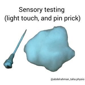
- Sensory testing of the lower limbs (light touch, and pin prick).
- Isometric muscle testing.
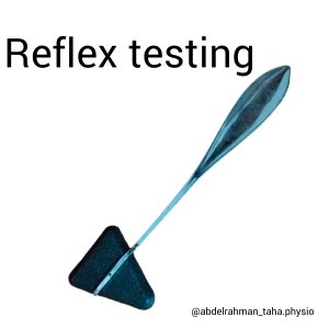
- Reflex testing.
- Neurodynamic testing.
These tests have been advised to demonstrate not only the presence of lumbosacral radiculopathy but also the degree of nerve root impairment (Bono and Garfin, 2004; De Luigi and Fitzpatrick, 2011; Petty, 2011a). Clinicians must perform a full neurological integrity test on any person who has neurological symptoms or spinally referred leg pain, not only to identify the presence of nerve root involvement but also to rule out serious spinal pathology such as cauda equina dysfunction (Bono and Garfin, 2004; Humphreys and Eck, 1999; Petty, 2011a; Refshauge and Gass, 2004).
Dermatomes (sensory testing)
Historically, dermatomal charts were created using a variety of methodologies for determining the sensory distribution of each nerve root (Foerster, 1933; Head and Canpbell, 1900; Keegan and Garrett, 1948; Sherrington, 1894), resulting in charts that were inconsistent between investigations.
One criticism levelled at older studies such as Head and Campbell (1900) and Foerster (1933) is that the procedures were not always precisely described. However, due to ethical considerations, repetition with previously used methods is not possible today. There is variation in how and where the nerve root is damaged, resulting in significant variation in dermatomal charts among people who appear to have the same nerve root involvement. Furthermore, dermatomes are known to overlap and vary between persons due to extra-dural anomalies in which two pairs of nerve roots may originate from a single dural sleeve or extra-dural anastomosis (Petty NJ and Moore AP. 2008).
Nitta et al. (1993) discovered significant diversity in distribution following a nerve root block in people with a herniated intervertebral disc. It is possible, however, that the diffusion of anaesthesia resulted in the anaesthesia of more than one nerve root. Lee et al. (2008) used several dermatomal charts to create a more accurate chart. There are no autonomous zones delineated due to level overlap, with the exception of the midline, which has negligible overlap. The diagnostic performance of sensory deficits, including hypoaesthesia, hypoalgesia, tingling, or numbness, has been studied in six studies (Albeck 1996; Kerr 1988; Knutsson 1961; Kosteljanetz 1984; Kosteljanetz 1988; Vroomen 2002 (primary care); Vucetic 1996). The forest plot has poor diagnostic performance in terms of sensitivity and specificity. Tawa et al. (2017) demonstrate that sensory testing of superficial soft touch and superficial pain is quite specific and might thus be used to rule out lumbo-sacral radiculopathy in individuals with low back and radiating leg symptoms.
Such variability makes being accurate about the nerve root level difficult based on dermatomal pattern alone, however simply identifying if a sensory loss has occurred in the leg for the lumbosacral nerve roots may be appropriate.
Myotomes (Motor tests)
Myotomes are motor supplies from a single nerve root. A radiculopathy is defined as weakened sets of muscles supplied by a single nerve root (Refshauge and Gass, 2004; Petty, 2011a).
Young et al. (1983) and Tsao et al. (2003) investigated the anatomical pattern of lumbosacral nerve root innervation of muscles in radiculopathy patients (identified surgically or radiologically). Tsao et al. (2003) performed both motor and sensory nerve conduction testing, whereas Young et al. (1983) only performed EMG. L5 was observed to preferentially innervate the peroneus longus (Tsao et al., 2003; Young et al., 1983), and S1 was observed to preferentially innervate the gastrocnemius (Tsao et al., 2003; Young et al., 1983) and hamstrings (Tsao et al., 2003). When compared to surgical findings, EMG findings incorrectly identified 16% of patients (Young et al., 1983), which may lower the robustness of the findings.
According to a review done by Van der Windt et al. (2011), paresis, or muscle weakness, performs poorly in diagnosing lumbar disc herniation. Kerr’s 1988 case-control research yielded more favourable results than this cohort study. When compared to surgical investigations, Vroomen 2002 (primary care) revealed a better specificity (0.93, 95% CI: 0.88 to 0.97) but a poor sensitivity (0.27, 95% CI: 0.20 to 0.37) of paresis. Also; When it comes to muscle wasting, Three studies in this review (Van der Windt et al. 2010) looked at muscular wasting and found comparable outcomes to those for muscle weakness (Albeck 1996; Kerr 1988; Knutsson 1961). Only Kerr (1988) outlined how muscle wasting was measured by calf circumference and offered a cut-off point for a positive test result (1 cm difference with the non-symptomatic side). Sensitivity ranged from 0.15 (Albeck 1996) to 0.38 (Kerr 1988), with specificity ranging from 0.50 (Knutsson 1961) to 0.94 (Kerr 1988). One case-control study (Kerr 1988) revealed very high specificity. Because of the small number of studies and the high degree of variability, the authors (Van der Windt et al. 2010) decided against statistical pooling of results.
Motor tests evaluated in the reviewed papers were largely functional tests of heel walk, heel raise, sit-to-stand, and resisted isometric contractions for hip flexion, knee extension, great toe extension, ankle dorsiflexion, and plantar flexion, according to Tawa et al. 2017. All studies used the same test to determine paresis, or muscle weakness. Sensitivity ranged from 0.13 (0.04-0.31) in the Bertilson et al. (2010) study to 0.61 (0.36-0.83) in the Suri et al. (2011) study. These findings have clinical implications in that motor tests are not ideal for screening out lumbo-sacral radiculopathy. Suri et al. (2011) showed the best specificity in this review for diagnosing S1 nerve root impingement. Bertilson (2010) offered a clear description of the actual execution of motor testing, allowing for duplication.
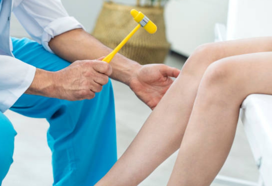
Deep tendon reflex testing
The patella reflex and the ankle jerk reflex are the most commonly evaluated tendon reflexes in the lower limb. These are thought to test the L3 or L4 nerve roots, as well as the S1 or S2 nerve roots (Jönsson and Strömqvist, 1996; Butler, 1991; Petty, 2011a). This stretch reflex is assumed to be reduced or missing when peripheral nerve routes (either afferent or efferent) are dysfunctional and increased when the central nervous system is dysfunctional. However, one study found a reduction in reflexes after injecting hypertonic saline into the zygapophyseal joints (Mooney and Robertson, 1976), suggesting that the appearance of a reduced reflex does not always indicate nerve root disease. The utility of reflexes in the diagnosis of nerve root compression is debatable in the literature.
Tawa et al. (2017) found that the diagnostic performance of reflex tests was notably good across studies, with specificity ranging from 0.60 (0.51-0.69) in the most recent Iversen et al. study to 0.93 (0.87–0.97) in the Vroomen (2002) study. The sensitivity, on the other hand, was moderate, with the highest value being 0.67 (0.21-0.94) in the Iversen (2013) study. As a result, deep tendon reflex tests should be used as confirmatory tests in the diagnosis of lumbosacral radiculopathy. However, the index test protocol and cut-off values for positivity were not supplied in several of the studies, and where they were provided, there were significant procedural variances.
Neurodynamic testing
The straight leg raise test has also been recommended as a method for detecting radiculopathy (Kobayashi et al., 2003). The test is known to have an effect on the lumbosacral nerve roots, resulting in excursion and strain (Gilbert et al., 2007 a and b, Goddard and Reid, 1965). The nerve root is pulled against the herniated disc by the positive test for radiculopathy, resulting in increased discomfort or neurological deficiency (Rabin et al., 2007).
Kobayashi et al. (2003) discovered a restriction in movement of the nerve root next to the disc herniation during SLR in 12 people with lumbosacral disc herniation, which considerably improved after the disc herniation was removed. Furthermore, blood flow measured with a laser Doppler flow metre was dramatically reduced at the angle where the people complained of pain during the SLR and significantly increased after discectomy. Another study by the same authors 7 years later in a larger cohort of 32 people validated the restriction and eventual improvement in nerve root excursion during the SLR (Kobayashi et al., 2010). Although sensitivity is often good to exceptional (Lauder et al., 2002; Poiraudeau et al., 2001; Rabin et al., 2007), specificity ranges from poor to fair (Lauder, 2002; Poiraudeau et al., 2001; Vroomen, 2002). The SLR is a neurodynamic test that assesses dysfunction in any of the neurological systems, from the nerve root to the continuation into the sciatic nerve and its distal connections (Butler, 1991; Butler, 2000; Shacklock, 2005a). As a result, its lack of specificity for radiculopathy is understandable, given that the test cannot detect lumbar disc herniations on its own. Individuals with radicular discomfort and radiculopathy are predicted to have a positive SLR; hence, the test is unable to distinguish between the two disorders.
According to Van der Windt et al. (2011), the Crossed Straight Leg Raising (XSLR) or crossed Lasègue’s test has a low sensitivity (varying between 0.23 and 0.43) but a good specificity (0.83 to 1.00). The Straight Leg Raising (SLR) or Lasègue’s test results also exhibited variation, with sensitivities ranging from 0.35 to 0.97 and specificities ranging from 0.10 to 1.00.
For the SLUMP test, two studies (Majlesi 2008; Stankovic 1999) published results. Stankovic (1999) presents the slump test results at various cut-off values (angles at which pain occurred), demonstrating that the slump test’s sensitivity was poor (0.44, 95% CI: 0.34 to 0.55) and specificity was slightly better (0.58, 95% CI: 0.28 to 0.85) when using a strict cut-off (pain radiating below the knee). When a gentler cut-off (pain anywhere) was used, sensitivity increased (and specificity fell). Majlesi (2008) reported a similar sensitivity (0.84) but greater specificity (0.83) for a positive test result using an unknown cut-off. The higher specificity may be due in part to the case-control design of this investigation, in which patients with back pain were chosen as controls if their MRI findings were absolutely normal.
concerning the diagnostic value of lower limb neurodynamic tests, such as the SLR test for the sacral plexus and the femoral nerve stretch test for the lumbar plexus. Tawa et al. (2017) discovered that the Trainor & Pinnington (2011) study had a good sensitivity and specificity of 1.00 (0.40–1.00) and 0.83 (0.52-0.98), respectively, with the remainder of the studies having low and intermediate diagnostic performance. Given these findings, lower limb neuro-dynamic tests (femoral nerve stretch test and SLR test) are more sensitive than specific, making them excellent for ruling out lumbo-sacral radiculopathy.
Validity of
Neurological Integrity Testing
Over a 9-month period, 5 extended scope physiotherapy practitioners assessed 123 patients who were then submitted for MRI scans in this study (Mercer and Smith, 2007). For a disc prolapse at the L3/4 level, the results showed a sensitivity of 100%, specificity of 98%, and accuracy of 100%; for L4/5, the results showed a sensitivity of 89%, specificity of 96%, and accuracy of 94%; and for L5/S1, the results showed a sensitivity of 93%, specificity of 85%, and accuracy of 89%. This study found that a comprehensive clinical examination, including neurological integrity, was an effective predictor of intervertebral disc herniation for experienced, highly educated physiotherapists. However, because these were all patients sent to specialty clinics with a higher risk of nerve root involvement, the population attending a typical physiotherapy clinic may not be representative of the general community. Furthermore, all physiotherapists had extensive training and were used to treating patients of this sort on a daily basis in their practice.
Most other investigations have not obtained such high sensitivity and specificity values (Iversen et al., 2013; Lauder et al., 2000; van der Windt et al., 2011). According to Lauder et al. (2000), the sensitivity and specificity of myotomal testing are 69% and 53%, respectively; reflex testing sensitivity is approximately 50% and specificity is around 90%; and sensory testing is 50% sensitive and 62% specific. When the results were merged, the specificity increased to 99% and the positive predictive value increased to 75%. Another intriguing discovery was that 90% of people with lumbosacral radiculopathy had at least one good neurological integrity finding. However, the gold standard employed was electrodiagnosis, which has only been shown to have a diagnostic accuracy of 54% (van Damme et al., 1979), lowering the confidence in the results. Van der Windt et al.’s 2011 systematic review found that the tests had poor validity, with sensitivity ranging from 0.27 to 0.62 and specificity ranging from 0.47 to 0.93. Reflex testing varied from 0.14 to 0.89 for sensitivity and 0.6 to 0.93 for specificity, whereas sensation testing ranged from 0.14 to 0.61 for sensitivity and 0.6 to 0.93 for specificity.
Such a large range and overall low validity do not support the use of these tests, but there were other limitations in both the systematic review (van der Windt et al., 2011) and original investigations that call the trustworthiness of this conclusion into question. To begin, each study’s methodology was so different, both in terms of the gold standard used to identify the exact level (primarily MRI or surgery; surgery may be more valid) and in determining whether the gold standard was evaluating nerve root compression or disc herniation. Other key diverse aspects were the methods employed to conduct the tests, with some studies missing details and others disclosing approaches that are not commonly used in practice.
One hundred and sixteen individuals were evaluated by both a physiotherapist and a medical practitioner, and the results were compared to MRI or CT scan findings (Iversen et al., 2013). Consideration of each individual test, as well as allowing for clinicians’ overall diagnosis of level at fault, did not provide sufficient evidence for the clinical use of these tests (likelihood ratios for clinicians’ diagnostic 1.74 (+ve) and 0.73 for L5 and 1.29 (+ve) and 0.61 for S1). The clinicians’ decision, however, was based on a very focused investigation and assessment of results that were not all well supported in the literature. Such clinical decision-making may not have accurately reflected current clinical practice, resulting in an incorrect diagnosis of nerve root involvement.
Takahashi et al. (1999) discovered a statistically significant increase in nerve root pressure (59.8 (+/- 50.3) mmHg compared to 15 (+/- 5.3) mmHg) among persons with MRI-diagnosed disc herniation and positive neurological integrity versus those with negative neurological integrity. The significance of this study lies in the fact that while all participants were diagnosed with disc herniation based on an MRI scan, not all had positive neurological integrity tests. It is also likely that the low sensitivity values reflect variances in individuals with a visibly recognised herniated disc, i.e., not all individuals with herniated discs have enough neurological damage to produce changes in neurological integrity, whereas some do. As a result, additional research is needed to back up these conclusions.
To recap, the research is generally not in favour of a single test for determining nerve root level but rather in favour of combining the tests and the clinical presentation of the patients (van der Windt et al., 2011; Tawa et al., 2017). Despite numerous studies indicating low validity measures for these tests, they are nonetheless recommended in clinical practice for assessing neurological integrity (Bono and Garfin, 2004; Humphreys and Eck, 1999; Petty, 2011a; Refshauge and Gass, 2004). Furthermore, due to the absence of alternative tests for the detection of nerve root involvement, these assays are still routinely used in practice.
References
- Albeck MJ. A critical assessment of clinical diagnosis of disc herniation in patients with monoradicular sciatica. Acta Neurochir (Wien) 1996;138:40-4.
- Bertilson BC, Brosjo E, Strender L. Assessment of nerve involvement in the lumbar spine: agreement between magnetic resonance imaging, physical examination and pain drawing findings. BMC Musculoskeletal Disord. 2010.11(202).
- Bono, M.B., Garfin, S.R. 2004 Spine. Philadelphia: Lippincott Williams and Wilkins.
- Butler, D. 2000. The Sensitive Nervous System Melbourne: NOI Press.
- Butler, D. 1991. Mobilisation of the Nervous System Melbourne: Churchill Livingstone.
- DeLuigi, A.J., Fitzpatrick, K.F. 2011 Physical examination in radiculopathy. Physical Medicine and Rehabilitation Clinics of North America 22:7-40.
- Deyo RA, Rainville J, Kent DL. What can the history and physical examination tell us about low back pain?. JAMA 1992;268(6):760-5.
- Foerster, O. 1933. The dermatomes in man. Brain. 1(56):1-39.
- Gilbert, K. K., Brismee, J.-M., Collins, D. L., James, R., Shah, R. V., Sawyer, S. F., Sizer, P. S. 2007a. Lumbosacral nerve root displacement and strain: part 1 a novel measurement technique during straight leg raise in unembalmed cadavers. Spine 32:1513-1520.
- Gilbert, K. K., Brismee, J.-M., Collins, D. L., James, R., Shah, R. V., Sawyer, S. F., Sizer, P. S. 2007b. Lumbosacral nerve root displacement and strain: part 2 straight leg raise conditions in unembalmed cadavers. Spine 32: 1521-1525.
- Goddard, M. D., Reid, J. D. 1965. Movements induced by straight leg raising in the lumbo-sacral roots, nerves and plexus, and in the intrapelvic section of the sciatic nerve. Journal of Neurology, Neurosurgery and Psychiatry 28: 12-18.
- Head, H., Campbell, A.W. 1900. The pathology of herpes zoster and its bearing on sensory localisation. Brain 23 (3): 353-362.
- Humphreys, S.C., Eck, J.C. 1999. Clinical evaluation and treatment options for herniated lumbar disc. American Family Physician. 59(3): 575-82.
- Iversen, T., Solberg, T.K., Romner, B., Wilsgaard, T., Nygaard, O., Waterloo, K., Brox, J.I., Ingebrigtsen, T. 2013. Accuracy of physical examination for chronic lumbar radiculopathy. BMC Musculoskeletal Disorders 14: 206.
- Jensen, M. C., Brant-Zawadzki, M. N., Obuchowski, N., Modic, M. T., Malkasian, D., & Ross, J. S. (1994). Magnetic resonance imaging of the lumbar spine in people without back pain. The New England journal of medicine, 331(2), 69–73.
- Jönsson, B., Strömqvist, B. 1996. Clinical characteristics of recurrent sciatica after lumbar discectomy. Spine. 21:500–505.
- Keegan, J.J., Garrett, F.D. 1948. The segmental distribution of the cutaneous nerves in the limbs of man. Anatomical Record 102: 409-37.
- Kerr RS, Cadoux-Hudson TA, Adams CB. The value of accurate clinical assessment in the surgical management of the lumbar disc protrusion. J Neurol Neurosurg Psychiatry 1988;51:169-73.
- Knutsson B. Comparative value of electromyographic, myelographic and clinical neurological examinations in diagnosis of lumbar root compression syndrome. Acta Orthop Scand Suppl 1961;49:1-134.
- Kobayashi, S., Takeno, K., Yayama, T., Awara, K., Miyazaki, T., Guerrero, A., Baba, H. 2010. Pathomechanisms of sciatica in lumbar disc herniation: effect of periradicular adhesive tissue on electrophysiological values by an intraoperative straight leg raising test. Spine 35(22): 2004-2014.
- Kobayashi, S., Shizu, N., Suzuki, Y., Asai, T., Yoshizawa, H. 2003. Changes in nerve root motion and intraradicular blood flow during an intraoperative straight leg raise test. Spine 28: 1427-1434.
- Kosteljanetz M, Espersen JO, Halaburt H, Miletic T. Predictive value of clinical and surgical findings in patients with lumbagosciatica. A prospective study (Part I). Acta Neurochir (Wien) 1984;73:67-76.
- Kosteljanetz M, Bang F, Schmidt-Olsen S. The clinical significance of straight-leg raising (Lasègue’s sign) in the diagnosis of prolapsed lumbar disc. Interobserver variation and correlation with surgical finding. Spine 1988;13:393-5.
- Lauder, T.D. 2002. Physical examination signs, clinical symptoms, and their relationship to electrodiagnostic findings and the presence of radiculopathy. Physical Medicine and Rehabilitation Clinics of North America 13(3):451-67.
- Lauder, T.D., Dillingham, T.R., Andary, M., Kumar, S., Pezzin, L.E., Stephens, R.T., Shannon S. 2000. Effect of history and exam in predicting electrodiagnostic outcome among patients with suspected lumbosacral radiculopathy. American Journal of Physical Medicine and Rehabilitation 79(1):60-8.
- Lee, M.W.L., McPhee, R.W., Stringer, M.D. 2008. An evidence based approach to human dermatomes. Clinical Anatomy 21:363-373.
- Majlesi J, Togay H, Unalan H, Toprak S. The sensitivity and specificity of the Slump and the Straight Leg Raising tests in patients with lumbar disc herniation. J Clin Rheumatol 2008;14:87-91.
- Mercer, C., Smith, T. 2007. Does the clinical impression of extended scope physiotherapists match the findings on MRI scans in people with low back pain? Paper presented at the World Physical Therapy Conference, Vancouver, 2-6th June.
- Mooney, V., Robertson, J. 1976. The facet syndrome. Clinical Orthopedics 115: 149-156.
- Nitta, H., Tajima,T., Sugiyama, H., Moriyama, A. 1993. Study on dermatomes by means of selective lumbar spinal nerve block. Spine, 18: 1782-86.
- Petty, N.J. 2011a. Neuromusculoskeletal Examination and Assessment: A Handbook for Therapists. London: Churchill Livingstone 4th edition.
- Petty NJ and Moore AP. Neuromusculoskeletal examination and assessment. A handbook for therapists. 5th Edition. Churchill Livingstone. 2008.
- Poiraudeau, S., Foltz, V., Drape, J.L., Fermanian, J., Lefevre-Colau, M.M., MayouxBenhamou, M.A., Revele, M. 2001.Value of the bell test and the hyperextension test for diagnosis in sciatica associated with disc herniation: comparison with Lasègue’s sign and the crossed Lasègue’s sign. Rheumatology 40: 460–6.
- Rabin, A., Gerszten, P.C., Karausky, P., Bunker, C.H., Potter, D.M., Welch, W.C. 2007. The sensitivity of the seated straight leg raise test compared with supine straight leg raise test in patients presenting with magnetic resonance imaging evidence of lumber nerve root compression. Archives of Physical Medicine and Rehabilitation 88: 840-843.
- Refshauge, K. Gass, E. 2004 Musculoskeletal Physiotherapy: Clinical Science and Evidence Based Practice 2nd edition Melbourne: Butterworth Heinemann.
- Scharfer A, Hall T, Briffa K. Classification of low back-related leg pain—A proposed patho-mechanism-based approach. Man Ther. 2007;14(2):222-30.
- Shacklock, M. 2005a. Clinical Neurodynamics: A New System of Musculoskeletal treatment Philadelphia: Elsevier.
- Sherrington, C.S. 1894. Experiments in examination of the peripheral distribution of fibres of the posterior roots of some spinal nerves. Philosophical Transactions of the Royal Society 184B: 641–763.
- Stankovic R, Johnell O, Maly P, Willner S. Use of lumbar extension, slump test, physical and neurological examination in the evaluation of patients with suspected herniated nucleus pulposus. A prospective clinical study. Man Ther 1999;4:25-32.
- Suri P, Rainville J, Katz JN, Jouve C, Hartigan C, Limke J, Pena E, Li L, Swaim B, Hunter DJ. The accuracy of the physical examination for the diagnosis of midlumbar and low lumbar nerve root impingement. Spine. 2011;36(1):63–73.
- Tawa, N., Rhoda, A., & Diener, I. (2017). Accuracy of clinical neurological examination in diagnosing lumbo-sacral radiculopathy: a systematic literature review. BMC musculoskeletal disorders, 18(1), 93.
- Takahashi, K., Shima, I., Porter, R.W. 1999. Nerve root pressures in lumbar disc herniation. Spine 24(19): 2003-2006.
- Trainor K, Pinnington M. Reliability and diagnostic validity of the slump knee bend neurodynamic test for upper/mid lumbar nerve root compression: a pilot study. Physiotherapy. 2011;97:59–64.
- Tsao, B.E., Levin, K.H., Bodner, R.A. 2003. Comparison of surgical and electrodiagnostic findings in single root lumbosacral radiculopathies. Muscle Nerve 27:60-64.
- van Damme, W., Hessels, G., Verhelst, M., van Laer, M., van Es. I. 1979. Relative efficiacy of clinical examination, electromyography, plain film radiography, myelography and lumbar phlebography in the diagnosis of low back pain and sciatica Neuroradiology 18: 109-118.
- van der Windt, D.A.W.M., Simons, E., Riphagen, I.I., Ammendolia, C., Verhagen, A.P., Laslett, M., Devillé, W., Deyo, R.A., Bouter, L.M., de Vet, H.C.W., Aertgeerts, B. 2011. Physical examination for lumbar radiculopathy due to disc herniation in patients with low-back pain (Review). The Cochrane Collaboration.
- Vroomen PCAJ, de Krom MCTFM, Wilmink JT, Kester ADM, Knottnerus JA. Diagnostic value of history and physical examination inpatients suspected of sciatica due to disc herniation: A systematic review. J Neurol. 1999;246:899–906.
- Vucetic N, Svensson O. Physical signs in lumbar disc hernia. Clin Orthop Relat Res 1996;333:192-201.
- Young, G., Getty, J., Jackson, A., Kirwan, E., Sullivan, M., Wynn Parry, C. 1983. Variations in the pattern of muscle innervation by the L5 and S1 nerve roots. Spine 8(6):616-624.