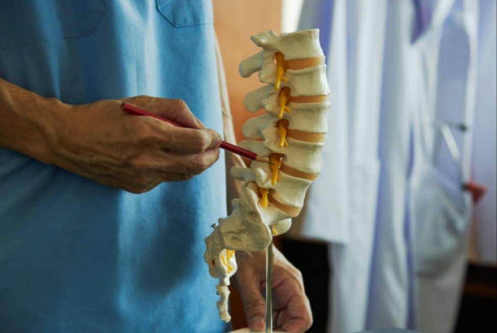
The nerve roots supplying the lumbosacral region originate from the lumbosacral part of the spinal cord and terminate in the conus medullaris, the cone-shaped lower end of the spinal cord. The tip of conus medullaris is often located at approximately the L1–2 intervertebral level, but it may end as high as T12–L1 or as low as L2–3. Therefore, the lower lumbar and sacral nerve roots run within the vertebral canal, where they are largely enclosed in the dural sac, before exiting the vertebral column. Such typical neuroanatomic features of the lumbosacral region have an important contribution to the natural course of lower back pain. In this article, I am going to share some insights that might assist your clinical reasoning for patients with lower back pain. Let’s start!
The long bundle of lumbosacral nerve roots, called the cauda equina, consists of the lumbar nerve roots that run along with the sacral and coccygeal nerve roots within the dural sac. The nerve roots of the cauda equina are bathed in cerebrospinal fluid (CSF).
The nerve fibers ‘within each nerve root’ are gathered into a single trunk, but near the spinal cord, they are separated into smaller bundles called rootlets. The rootlets of each ventral root attach to the ventrolateral aspect of the cord, while those of the dorsal roots attach to the dorsolateral sulcus of the cord. Proceeding caudally, a sensory dorsal and motor ventral nerve root come together in the lateral region of the dural sac, finally departing from the dural sac via the dural root sleeve which contains an extension of the subarachnoid space.
At the termination of the root sleeve, the roots divide into a number of fascicles. The sensory root fascicles merge into the dorsal root ganglion, and the motor fascicles blend in distally to the dorsal root ganglion as the mixed spinal nerve is formed, the ventral ramus of which goes on to become part of the lumbosacral plexus. Thus, the nerve roots are sheathed with pia mater and bathed in CSF as far as the spinal nerve.
Each exiting lumbar nerve root is numbered according to the vertebra under whose pedicle it enters the lumbar intervertebral foramen.
Each intervertebral disc is anatomically related to two nerve roots, and each emerging nerve root and nerve pass two intervertebral discs.
An unusually high or low position of the dural end-sac will lead to a similarly high or low departure of the root sleeves relative to the disc. This relationship affects the mobility of the intradural nerve root at the level of the intervertebral disc, and thus the liability of the nerve root to compression at various spinal disc levels.
The lumbar intervertebral foramina (IVFs) form a series of canalized tunnels through which the lumbar spinal nerve root emerges from the spinal canal.
At the lumbar IVF, the orifice has two parts:
1. superior and rigid part, where nerve components pass.
2. inferior part is more mobile and exposed to disc changes.
The ligamentous structures in the lumbar IVFs called the transforaminal ligaments (TFLs), which divided TFLs into three categories: internal ligaments, intraforaminal ligaments, and external ligaments. As about 50 to 53% of the foramen area is occupied by the nerve root, the presence of the TFL diminishes the superoinferior dimension to 35% of the available space of the IVF. Nevertheless, there is no evidence in the literature that those ligaments exert any pressure on the vessels transversing the foramen.
The greater the collapse of the intervertebral disc, the greater the reduction in foraminal height. So, the passage of nerve components in the bony upper part of the foramen protects the root somewhat from disc protrusion, facet joint osteoarthritis, and ascending of the inferior articular process during extension movements ‘as long as the height of the intervertebral space remains satisfactory’. Also, the dimensions of the lumbar intervertebral foramina show changes in features of IVFs with increasing age, height, and weight. So, determining these IVF components of spinal units provides a more dynamic assessment and evaluation of changes in the IVFs secondary to therapeutic interventions.
Let’s take a look at the lumbar lateral recess. Two levels can be distinguished within the lumbar lateral recess:
1. Ligamentous lateral recess:
At the disc level, formed by the lateral spinal canal is bordered ventrally by the annulus fibrosus and dorsolaterally by the facet joint capsule and the flaval ligament, containing the traversing nerve root.
2. Bony lateral recess:
At the pedicular level, which contains the root sleeve, is bordered ventrally by the vertebral body, laterally by the pedicle, and dorsolaterally by the base of the superior articular process.
Functionally, the anatomical distinction between two levels of the lateral recess is of clinical relevance. As, at the level of the ligamentous lateral recess, soft tissue changes, often posture-related, can compress the traversing nerve root or root sleeve more easily.
Finally, let’s take a brief overview of dorsal root ganglia (DRG) and variations in its position.
There are three positions for DRG, which are:
A. spinal canal type:
The proximal end of the DRG is located at the medial edge of the pedicle.
B. intraforaminal type:
The proximal end of the DRG is located between the center and medial edge of the pedicle.
C. extraforaminal type:
The proximal end of the DRG is located lateral to the center of the pedicle.
Lumbar DRG motion is dynamic with slight positional change with activity. Also, DRG is positioned more lateral from L1 through L5.
DRG is very sensitive to mechanical compression and is closely related to abnormal sensation and radiating pain (it is able to generate its own action potentials), as when a nerve root is compressed it won’t cause pain unless it has already been inflamed. But when a dorsal root ganglion is compressed it will fire off a lot of nociceptive action potentials, even in a perfectly healthy state.
I hope that was interesting for you. Thank you for reading this article. If you find this interesting, please share it with someone who might benefit from it!
References:
1. Church CP, Buehler MT (1991) Radiographic evaluation of the corporotransverse ligament at the L5 intervertebral foramen: a cadaveric study. J Manipulative Physiol Ther 14:240-248 .
2. Kostelic J, Haughton VM, Sether L (1992) Proximal lumbar spinal nerves in axial MR imaging, CT, and anatomic sections.Radiology 183(1):239 .
3. Kostelic JK, Haughton VM, Sether LA (1991) Lumbar spinal nerves in the neural foramen: MR appearance. Radiology 178(3):837.
4. Wiltse LL (2000) Anatomy of the extradural compartments of the lumbar spinal canal. Peridural membrane and circumneural sheath. Radiol Clin North Am 38(6):1177.
5. Louis R. Topographic relationship of the vertebral column, spinal cord, and nerve roots.Anat Clin.1978;1:3–12.
6. Park HK, Rudrappa S, Dujovny M, Diaz FG. Intervertebral foraminal ligaments of the lumbar spine: anatomy and biomechanics. Childs Nerv Syst. 2001 Apr;17(4-5):275-82.
7. Min JH, Kang SH, Lee JB, Cho TH, Suh JG. Anatomic analysis of the transforaminal ligament in the lumbar intervertebral foramen. Neurosurgery. 2005 Jul;57(1 Suppl):37-41; discussion 37-41.
