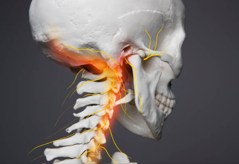
The neck has anatomic structures that are involved in balance control (cervical afferents), vascular tone regulation (carotid sinuses), blood circulation (carotid and vertebral arteries), and mechanical support of the cervical spinal cord. Therefore, the symptoms such as dizziness, imbalance, or vertigo associated with cervicocephalic movements could also be attributed to vestibular (inner ear), visual, vascular, neurovascular, cervicoproprioceptive, or cervical spinal cord dysfunction. In this article, I am going to share some neuroanatomical insights that might assist your clinical reasoning for patients with neck pain. Let’s get right into it!
The union of ventral (motor) and dorsal (sensory) roots that emerge bilaterally from the cervical cord leads to the formation of cervical nerves (mixed spinal nerves). In other words, the posterior and anterior spinal nerve roots that emerge from the cervical spinal cord combine together to form a mixed nerve before entering the cervical intervertebral foramen. The nerve root fibers then pass obliquely through the intervertebral foramen and down the foraminal groove. Upon emerging from the intervertebral foramen, the mixed nerve divides into two rami: the posterior and anterior rami. Within the dural sac, the upper cervical nerve roots tend to pass upward toward the spinal nerve. Lower cervical nerve roots pass transversely or slightly downward. In general, the nerves are aligned almost horizontally, so any symptoms from impingement on a nerve root by a cervical disc displacement will most likely be monoradicular.
The nerve roots from C1 to C7 are named after the vertebral body below, whilst the C8 nerve root exits below the C7 vertebral body, and the nerve roots below this are named after the vertebral body above.
The cervical nerve root normally occupies only one-fourth to one-fifth of the intervertebral foramen. From the intradural to extra vertebral region, the nerve roots must pass through the following three successive areas:
Medial area:
A relatively large amount of space. Within the medial orifice of the intervertebral foramen. Ligamentum flavum forms its posterior wall.
Intermediate area:
Narrow area. Between the uncus in front and posterior facet joint behind, with the pedicle below, constitutes the intervertebral foramen.
Lateral area:
Involved with the spinal nerve, rests in the furrow of the transverse process. The bottom of this transverse furrow is perforated by the vertebral foramen, providing passage to the vertebral artery. This is an essential relation to the cervical nerve, as vertebral artery wall defect is an indirect cause of radiation &/or radiculopathy.
Finally, these three areas are aligned on an axis oriented at 45° forward compared to the sagittal plane. Because of this oblique angle, the bony borders of the cervical intervertebral foramen are visible only on oblique MRI & X-ray.
The adult cervical disc comprises four distinct structures:
1) a crescent-shaped anterior annulus fibrosus, thick anteriorly and tapered laterally toward the uncinate process;
2) the central fibrocartilaginous core called as nucleus pulposus;
3) periosteofascial tissue overlying the uncovertebral area;
4) a thin posterior annulus fibrosus. This is bordered anteriorly by the median anterior longitudinal ligament and posteriorly by the broad posterior longitudinal ligament with median fibers running inferior-superior, and alar fibers running at 45° covering the postero-lateral aspect of the disc.
There are anatomic relationships between the nerve roots and the discs in the intervertebral foramen of the cervical spine:
1. “shoulder relationship”: when the disc is proximal to the nerve root.
2. “anterior relationship”: when the disc is located just anteriorly to the nerve root.
3. “axillary relationship”: when the disc is distal to the nerve root.
4. “no contact relationship”: when the disc does not have contact with the nerve root.
These anatomical relationships may give some reasoning about the complex nerve root and disc relationships, the high incidence of nerve root compression at C5–C6 space (for C6 nerve root) and C6–C7 space (for C7 nerve root), why C8 radicular symptoms and/or radiculopathy are so rare (as previously mentioned), and the mechanisms behind painless radiculopathy.
Also, it is interesting to note that although the widths of the C5 and C6 roots are often wider than those of C7 and C8, the C7 root is the most commonly involved, followed by the C5, C6, and C8 roots. Therefore, while investigating the causes of cervical radiculopathy, one should consider not only the width of the nerve root but also the relationship to the surrounding bony anatomy.
Furthermore, the cervical region has;
1. a posterior longitudinal ligament, a double-layered ligament that spans the entire posterior portion of the vertebral body.
2. posterior portion of the annulus, which is broader and firmer.
3. uncovertebral joints of von Luschkn.
All these factors protect the cervical nerve roots and the spinal cord from the protruding disc material and do not favor the herniation of disc material as a common source of nerve root pressure. Therefore, cervical nerve root compression from disc herniation is not so frequent. In order to contact the nerve, the disc must herniate into the intervertebral foramen and thus protrude in a dorsolateral direction. This direction of disc herniation is prevented or at least minimized in the cervical region. However, understanding the relationships within the intervertebral foramen should not be based on these assumptions.
Therefore, current treatment strategies are devised in line with the symptoms and the mechanical responses to anatomic changes, not simply following a pathoanatomical concept.
I hope that was interesting to you. Thank you for reading this article. If you find this interesting, please share it with someone who might benefit from it!
Reference:
1. Kobayashi R, Iizuka H, Nishinome M, Iizuka Y, Yorifuji H, Takagishi K. A cadaveric study of the cervical nerve roots and spinal segments. Eur Spine J. 2015 Dec;24(12):2828-31.
2. Tanaka N, Fujimoto Y, An HS, Ikuta Y, Yasuda M. The anatomic relation among the nerve roots, intervertebral foramina, and intervertebral discs of the cervical spine. Spine (Phila Pa 1976). 2000 Feb 1;25(3):286-91.
3. Goldstein B. Anatomic issues related to cervical and lumbosacral radiculopathy. Phys Med Rehabil Clin N Am. 2002 Aug;13(3):423-37.
4. Demondion X, Lefebvre G, Fisch O, Vandenbussche L, Cepparo J, Balbi V. Radiographic anatomy of the intervertebral cervical and lumbar foramina (vessels and variants). Diagn Interv Imaging. 2012 Sep;93(9):690-697.