
Introduction:
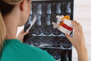
Low back pain (LBP) is a complex illness that includes physiological, psychological, and brain alterations (Nijs J. et al. 2017). LBP is a complex condition that includes physiological, psychological, and neurological alterations (Nijs J. et al. 2017). Many researchers in the field of LBP, including physicians, believe that choosing the most effective treatment for the individual patient is impossible without a deeper understanding of the biological component of the biopsychosocial model (Hancock MJ et al. 2011). The intervertebral disc is the most common source of persistent nociception in LBP, accounting for at least 39% of cases (Schwarzer AC et al. 1995). Magnetic resonance imaging images of outer annulus disturbances as high-intensity zones correlate with the induction of familiar pain during discography (Rankine JJ, et al. 1999; Saifuddin A, et al. 1998; Aprill C, and Bogduk N, et al. 1992; Sachs BL, et al. 1987), However, not all discs that cause pain during discography have aberrant magnetic resonance imaging signal intensity, disc contour, or severe symptoms of degeneration (Milette PC, et al. 1999).
Discogenic low back pain pathomechanisms (Ohtori S. et al., 2018):
- Innervation: An animal model and human specimens indicated sensory nerve innervation of lumbar intervertebral discs (IVDs) as well as sensory nerve ingrowth into the inner layer of the deteriorated IVD (deep nerve ingrowth).
- Inflammation: many proinflammatory chemicals have been found by researchers.
- Hypermobility: In deteriorated IVD, hypermobility of the motion segment is common.
These are regarded to be the primary causes of discogenic low back pain.
Mechanically reducible discogenic back pain diagnosis:
Provocation discography (Aprill CN, 1997; Guyer RD, et al, 2003; Schwarzer AC, et al, 1995) is a way of directly confronting the disc as a diagnostic sign of discogenic pain. It could be used as a reference standard to measure the diagnostic accuracy of clinical factors. However, because this test is intrusive and costly, it should not be administered to all individuals. As a result, evidence-based clinical diagnosis with adequate accuracy will eliminate the need for intrusive or costly diagnostic procedures.
Centralization has sensitivity, specificity, and positive probability ratios of 0.92, 0.64, and 2.5, respectively, while peripheralization has sensitivity, specificity, and positive likelihood ratios of 0.69, 0.64, and 1.9, respectively. The sensitivity, specificity, and positive probability ratios of these two indications are 0.92, 0.52, and 1.96, respectively (Bogduk N, Lord S, 1997). The centralization phenomenon (CP) (McKenzie RA, 1981) was discovered to be highly specific (80–100%) to concordant pain provocation during discography but with low sensitivity (35–45%) depending on patient distress and other circumstances (Laslett M. et al., 2005). Only during a repetitive movement assessment, which requires patients to complete standardised repeated test motions, can the CP be evoked (McKenzie RA and May S. 2003). If a patient is unable to finish this evaluation, additional clinical tests are required to determine whether a concordant pain response to discography is likely.
In a 2006 study, Laslett M. and colleagues discovered that a history of persistent pain between acute episodes, a significant loss of extension, and a subjective report of ‘vulnerability’ in what is known as the ‘neutral zone’ had specificities of 83–92% and likelihood ratios ranging from 2.0 to 4.1. Although two variable combinations were highly specific to positive discographies, no suitable screening tool for ruling out positive discographies was found. Three clinical variables have low predictive potential for lumbar discography outcomes, whereas two combinations of variables are highly specific for positive discography. Furthermore, Levi D. et al. (2018) discovered that a positive history of severe episodic low back pain may be a strong signal for a discogenic aetiology. The diagnostic confidence odds are 18.2, with a 95% confidence level. Young S. and colleagues discovered in a 2003 study that patients who experience midline lumbar discomfort and pain when rising from a sitting position are more likely to have discogenic pain.
The clinical reasoning approach utilised by clinicians is characterised by the formation of a diagnosis based on a combination of findings. To do this, I proposed a set of characteristics that are highly diagnostic of the discogenic origin of back pain presentation. These variables are:
- Patients with lumbar discomfort in the midline.
- Pain when rising from a seated position.
- A history of chronic pain between acute bouts.
- Significant reduction in extension ROM.
- Subjective report of ‘vulnerability’ in the so-called ‘neutral zone”.
- The presence of the phenomenon of centralization.
References:
- Aprill C, Bogduk N. High-intensity zone: a diagnostic sign of painful disc on magnetic resonance imaging. Br J Radiol 1992;65:361–9.
- Aprill CN (1997) Diagnostic disc injection. II Diagnostic lumbar disc injection. In: Frymoyer JW, Ducker TB, Hadler NM, Kostuik JP, Weinstein JN, Whitecloud TS (eds) The adult spine: principles and practice, 2nd edn. Lippincott-Raven Publishers, Philadelphia, pp 539–562.
- Bogduk N, Lord S. A prospective study of centralization of lumbar and referred pain: a predictor of symptomatic disc and annular competence: commentary. Pain Med J Club 1997;3:246–8.
- Guyer RD, Ohnmeiss DD, Vaccaro A (2003) Lumbar discography. Spine J 3:11–27.
- Hancock MJ, Maher CG, Laslett M, Hay E, Koes B. Discussion paper: what happened to the ‘bio’ in the bio-psycho-social model of low back pain? Eur Spine J. 2011;20(12):2105–10.
- Laslett M, O berg B, Aprill CN, McDonald B (2005) Centralization as a predictor of provocation discography results in chronic low back pain, and the influence of disability and distress on diagnostic power. Spine J 5:370–380.
- Laslett M, Aprill CN, McDonald B, Oberg B. Clinical predictors of lumbar provocation discography: a study of clinical predictors of lumbar provocation discography. Eur Spine J. 2006 Oct;15(10):1473-84.
- Levi D, Carnahan D, Horn S, Levin J. Is a History of Severe Episodic Low Back Pain an Indicator of a Discogenic Etiology? Pain Med. 2018 Jul 1;19(7):1334-1339.
- McKenzie RA (1981) The lumbar spine: mechanical diagnosis and therapy. Spinal Publications Ltd, Waikanae, pp 22–23.
- McKenzie RA, May S (2003) Mechanical diagnosis and therapy: the lumbar spine, 2nd edn. Spinal Publication New Zealand Ltd, Waikanae, pp 404–426.
- Milette PC, Fontaine S, Lepanto L, et al. Differentiating lumbar disc protrusions, disc bulges, and discs with normal contour but abnormal signal intensity. Magnetic resonance imaging with discographic correlations. Spine 1999;24:44–53.
- Nijs J, Clark J, Malfliet A, et al. In the spine or in the brain? Recent advances in pain neuroscience applied in the intervention for low back pain. Clin Exp Rheumatol. 2017; 35 Suppl 107 (5): 108–15.
- Ohtori S, Miyagi M, Inoue G. Sensory nerve ingrowth, cytokines, and instability of discogenic low back pain: A review. Spine Surg Relat Res. 2018 Jan 27;2(1):11-17.
- Rankine JJ, Gill KP, Hutchinson CE, et al. The clinical significance of the high-intensity zone on lumbar spine magnetic resonance imaging. Spine 1999;24:1913–9.
- Sachs BL, Vanharanta H, Spivey MA, et al. Dallas discogram description: a new classification of CT/discography in low-back disorders. Spine 1987;12:287–94.
- Saifuddin A, Braithwaite I, White J, et al. The value of lumbar spine magnetic resonance imaging in the demonstration of anular tears. Spine. 1998;23:453–7.
- Schwarzer AC, Aprill C, Derby R, Fortin JD, Kine G, Bogduk N. The prevalence and clinical features of internal disc disruption in patients with chronic low back pain. Spine 1995;20:1878–83.
- Young S, Aprill C, Laslett M. Correlation of clinical examination characteristics with three sources of chronic low back pain. Spine J. 2003 Nov-Dec;3(6):460-5.