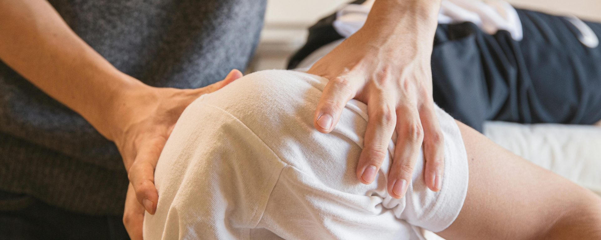
Clinician generated forces refer to the external forces exerted by the clinician on a patient’s body segment to diagnose, treat, and/or to facilitate recovery.
Clinician generated forces can be exerted through various techniques, such as joint glides, thrusts, or the use of mechanical devices to assist in the diagnosis, treatment, and rehabilitation of various conditions. For example, sustained pressure or repetitive joint glides may help with assessing the restriction of a joint movement, while thrust techniques that involve a quick, controlled application of a force to a joint may help with restoring movement.
Such external forces exerted by the clinicians on a human body segment as an intervention are called as ‘clinician generated forces’.
Patient generated forces, in contrast, refer to the internal forces produced by the patient through their own voluntary actions or physiological processes. These forces include voluntary movements such as pushing or pulling, breathing, and the body’s inherent / involuntary ability to self-correct.
The effectiveness of clinician-generated forces often depends on the understanding of biomechanics and the specific needs of the patient. By applying the right amount of force at the appropriate angle and duration, clinicians can optimize diagnostic and/or treatment outcomes. Additionally, these forces can be combined with patient generated forces—where patients actively engage in movements—to create a comprehensive rehabilitation strategy that addresses both external and internal dynamics of recovery.
Examples of Clinician-Generated Forces:
I. Diagnostic Tests: Diagnostic manual tests like the Anterior Drawer Test are examples of how clinicians use controlled external forces to assess the structural integrity and function of joints and tissues. During the Anterior Drawer Test of the knee joint, the clinician applies an anterior force to the tibia while stabilizing the femur. This force is used to assess the anterior translation of the tibia in relation to the femur, which can indicate the integrity of the anterior cruciate ligament (ACL). The force applied is controlled and specific, aiming to replicate the stress that might cause symptoms or indicate a pathology. The clinician’s understanding and skill in applying the appropriate amount of force are crucial to the test’s accuracy and safety. An abnormal amount of movement or a lack of a firm endpoint may indicate an ACL tear or injury.
II. Manual Therapy: In manipulative therapy techniques such as joint glides or thrusts, the clinician applies specific forces to a patient’s joints and tissues. For example, during a spinal manipulation, the clinician uses their hands to apply a quick, controlled force to the spine to improve mobility and reduce pain.
High velocity low amplitude (HVLA) thrust technique is an example of clinician-generated force, with the hypothesis being that torque is created to separate the articular surfaces of joints. The kinematics of HVLA thrust in the cervical spine are not fully understood. However, typical parameters include:
Mean thrust duration: 158 ms, ranging from 117 to 250 ms (1) (2)
Mean de-rotation displacement before thrust: 4.8 degrees (1) (3)
Mean thrust displacement/angle: 11.4 degrees, ranging from 6.0 to 22.5 degrees (1)
Average peak thrust velocity: 127 degrees/sec (1)
Mean peak force applied: 118 N (4)
Facet gap increase: 0.9 ± 0.40 mm (5)
Mean rate of facet gapping: 6.2 ± 3.9 mm/s (5)
Peak force: 65 ± 4 N (5)
Rate of force: 440 ± 58 N/s (5)
III. Stretching Techniques: When performing passive stretching, the clinician applies a gentle, sustained force to stretch a patient’s muscles or tendons. This helps increase flexibility and range of motion.
For instance, the clinician might passively stretch the anterior compartment of thigh (rectus femoris and fascia lata) by holding the patient’s leg and applying a force to lengthen the muscle. In a study involving 7 high school male elite rugby players, the stretching force applied (for 10 min per session for 11 sessions) ranging from 124 to 164 N resulted in a 1 cm increase of myofascial length (6).
On the other hand, an acute bout of sub-maximal or maximal intensity of stretching (up to 50%, 75%, or 100% of the point of discomfort) force applied to quadriceps, hamstrings, and plantar flexors can impair the performance of drop jumps, squat jumps, and countermovement jumps (7).
IV. Resistance Training: In resistance exercises, the clinician may use external weights or resistance bands or use their own hands to apply force against the patient’s muscles. This helps strengthen specific muscle groups. The clinician may adjust the level of resistance to match the patient’s strength and rehabilitation goals.
V. Assistive Devices: Clinicians may use devices such as braces or orthoses to apply external forces that support, align, or correct a patient’s body position. For example, a knee brace can provide support and limit movement to aid in the healing of an injured knee.
Reference:
(1) Ngan JM, Chow DH, Holmes AD. The kinematics and intra- and inter-therapist consistencies of lower cervical rotational manipulation. Med Eng Phys. 2005 Jun;27(5):395-401
(2) Reed WR, Cao DY, Long CR, Kawchuk GN, Pickar JG. Relationship between Biomechanical Characteristics of Spinal Manipulation and Neural Responses in an Animal Model: Effect of Linear Control of Thrust Displacement versus Force, Thrust Amplitude, Thrust Duration, and Thrust Rate. Evid Based Complement Alternat Med. 2013;2013:49203
(3) Evans, David & Breen, Alan. . A Biomechanical Model for Mechanically Efficient Cavitation Production During Spinal Manipulation: Prethrust Position and the Neutral Zone. Journal of manipulative and physiological therapeutics.(2006);29. 72-82.
(4) Kawchuk GN, Herzog W, Hasler EM. Forces generated during spinal manipulative therapy of the cervical spine: a pilot study. J Manipulative Physiol Ther. 1992 Jun;15(5):275-8.
(5) Anderst WJ, Gale T, LeVasseur C, Raj S, Gongaware K, Schneider M. Intervertebral kinematics of the cervical spine before, during, and after high-velocity low-amplitude manipulation. Spine J. 2018
(6) Germain F, Lemarchand E, Perrin R. Sensory regulation and mechanical effects of sustained high intensity stretching of the anterior compartment of the thigh. J Bodyw Mov Ther. 2020 Apr;24(2):18-25. doi: 10.1016/j.jbmt.2020.02.028. Epub 2020 Feb 27. PMID: 32507143.
(7) Behm, D.G., Kibele, A. Effects of differing intensities of static stretching on jump performance. Eur J Appl Physiol 101, 587–594 (2007). https://doi.org/10.1007/s00421-007-0533-5