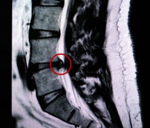
Determining whether a specific lesion is responsible for a patient’s symptoms is crucial when selecting candidates for potential spinal surgery. However, there isn’t always a direct correlation between radicular pain and lumbar disc herniation. One can exist without the other, making diagnosis complex. Both mechanical compression and inflammatory factors contribute to the pathogenesis of sciatica. Additionally, non-radicular or pseudoradicular pain can originate from the hip, sacroiliac joint, or spinal facets and may misleadingly present as pain in the lower extremity. Moreover, intermittent claudication from arterial occlusive disease can be confused with neurogenic claudication from cauda equina compression in spinal stenosis.
Historical studies highlight the complexity of these diagnoses. Hitselberger and Witten (1968) found abnormal lumbar and cervical myelographic findings in 110 out of 300 patients without symptoms of nerve root compression. Wiesel et al. (1984) reported disc herniations in 35% of asymptomatic individuals in their CT studies. Similarly, Boden et al. (1990) found herniated discs or spinal stenosis in one-third of asymptomatic individuals, with stenosis incidence increasing with age. In a long-term study, Borenstein et al. (2001) concluded that asymptomatic disc herniations did not increase the risk of developing low back pain. Conversely, Modic et al. (1995) observed normal lumbar spinal MRIs in some patients with acute lumbar radiculopathy symptoms.

Interestingly, disc herniation can sometimes appear on the side opposite to clinical symptoms (Safdarian M, et al. 2016; Ruschel LG, et al. 2021), making it a chance finding unrelated to the patient’s pain. An unwarranted decision to operate can result in removing an asymptomatic disc herniation while the true cause of pain remains untreated.
In summary, no single feature can reliably distinguish a symptomatic disc herniation from an incidental finding. This highlights the importance of a comprehensive and cautious approach in diagnosing and treating lower back pain and related symptoms. For more insights into the relationship between MRI findings and presenting symptoms in low back pain patients, check out my recent blog posts (1, & 2).
Stay informed and approach each case with the thoroughness it deserves.
References:
Boden SD, Davis DO, Dina TS et al (1990) Abnormal magneticresonance scans of the lumbar spine in asymptomatic subjects.
Borenstein DG, O’Mara JW Jr, Boden SD et al (2001) The value of magnetic resonance imaging of the lumbar spine to predict low-back pain in asymptomatic subjects: a seven-year follow-up study. J Bone Joint Surg Am 83-A(9):1306.
Hitselberger WE, Witten RM (1968) Abnormal myelograms in asymptomatic patients. J Neurosurg 28(3):204.
Modic MT, Ross JS, Obuchowski NA et al (1995) Contrastenhanced MR imaging in acute lumbar radiculopathy: a pilot study of the natural history. Radiology 195(2):429.
Wiesel SW, Tsourmas N, Feffer HL et al (1984) A study of computer-assisted tomography. I. The incidence of positive CAT scans in an asymptomatic group of patients. Spine 9(6):549.
Ruschel LG, Agnoletto GJ, Aragão A, Duarte JS, de Oliveira MF, Teles AR. Lumbar disc herniation with contralateral radiculopathy: a systematic review on pathophysiology and surgical strategies. Neurosurg Rev. 2021 Apr;44(2):1071-1081.
Safdarian M, Farzaneh F, Rahimi-Movaghar V. Contralateral Radiculopathy: A Kernohan-Woltman Notch-like Phenomenon. Asian J Neurosurg. 2018 Jan-Mar;13(1):165-167.
