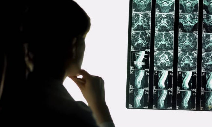
Role of MRI in spine physical therapy practice.

Magnetic resonance imaging (MRI) detected lumbar pathology may be a significant factor in the context of low back pain (LBP) recurrence (M Hildebrandt et al, 2017). Investigating lumbar pathology with MRI, a non-invasive technique is a standard practice in medicine (Milette et al, 1999). Since MRI imaging reveals anatomical and morphologic features of the spine, the results do not directly determine the cause of pain. … Continue reading Role of MRI in spine physical therapy practice.
Kernig’s sign

Purpose of Kernig’s sign: To determine if the patient has inflammation of the meninges (meningitis) (1). Patient position: Lying supine (conventional practice). VM Kernig originally elicited this sign by having the patient in sitting position. Examiner position: Standing beside the bed, facing the patient. Procedure: With the patient lying supine, flex one of the patient’s legs (hip and knee joints) to 90 degrees, and then … Continue reading Kernig’s sign

Brudziński’s symphyseal sign

Purpose of Brudziński’s symphyseal sign : To determine if the patient has inflammation of the meninges (meningitis) (1). Patient position: Lying supine. Examiner position: Standing beside the bed, facing the patient. Procedure: With the patient lying supine, place the heel of your hands on patient’s pubic symphysis and apply gentle pressure. Outcome: If this manoeuvre causes involuntary flexion of the patient’s hips and knees and … Continue reading Brudziński’s symphyseal sign

Cervical adherent nerve root dysfunction: classification and therapy – A case report

Introduction: Mechanical diagnosis and therapy (MDT) of the cervical spine, also known as the McKenzie method, has been the focus of research in the field of musculoskeletal physical therapy in recent years. The MDT focuses on the patient’s symptomatic and mechanical responses to the systematic application of repetitive end-range movements. Adherent nerve root (ANR) is a specific form of dysfunction that occurs secondary to trauma, … Continue reading Cervical adherent nerve root dysfunction: classification and therapy – A case report

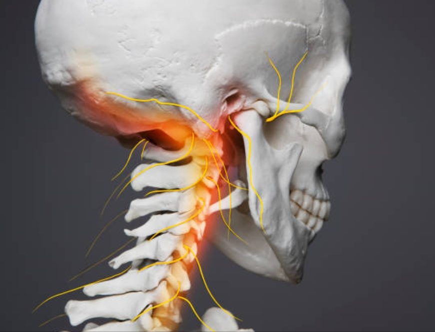
Journey into the upper cervical segments | A primer on neuroanatomy.

The upper two cervical segments formed by atlanto-occiptal & atlanto-axial joints are anatomically dissimilar from the functional units below. In this article, I am going to share some insights that might assist your clinical reasoning for patients with upper cervical neck pain. The craniocervical junction (CCJ) is a complex transitional region between the base of the skull and the upper cervical spine. It is formed … Continue reading Journey into the upper cervical segments | A primer on neuroanatomy.